The mission of the NWA is to globally promote nanotechnology solutions adoption across industries by connecting entrepreneurs with researchers, start-ups with investors, providers with potential customers, employers with job seekers, key players with one another, in an independent and mainly industry-oriented advocacy group – inclusive of business, academia, business-supporting associates, as well as affiliate government agencies and other associations.
Biologically driven design leads to the development of novel multi-functional materials, miniaturized electromechanical systems, and reliable living tissues as a more sustainable solution to pressing technological problems facing the human race.
Credit: Edited by: Esmaiel Jabbari (University of South Carolina, USA), Deok-Ho Kim (University of Washington, USA), Luke P Lee (University of California, Berkeley, USA), Amir Ghaemmaghami (University of Nottingham, UK), Ali Khademhosseini (Harvard University, USA & Massachusetts Institute of Technology, USA)
How should we respond to technologies around us that are inefficient, wasteful, pollute our environment, and overburden our health care system? In their latest three three volume "Handbook of Biomimetics and Bioinspiration" published with World Scientific, renowned researcher Dr Jabbari and his co-editors provide answers to this question. Nature has evolved over millions of years to create structures with amazing complexity. Nowhere this complexity is more apparent than in plants and animals: mimicking non-sticking of lotus leaves to produce self-cleaning devices, stickiness of gecko's feet for space walking, water strider's walking on water to build aquatic reconnaissance microrobots, mimicking laminated structure of the skin to generate advanced membranes for water purification, anti-reflectivity of cicada's wings to build improve medical imaging systems, mimicking self-healing of earthworms to make tires that repair themselves, desert beetle's water collecting ability to design water storage systems in dry and hot climates, and mimicking uneven wetting of butterfly wings to trigger motion in a particular direction. Nature-driven design that mimics the hierarchical complexity of biological systems leads to the development of more reliable miniaturized and compact devices that perform multiple tasks. These include muscle mimetic actuators and robots, algae mimetic photoreception devices, sea star mimetic light sensing, ocellus mimetic optical devices, cell membrane mimetic nanochannels, nerve tissue mimetic sensory systems, olfactory mimetic odor sensing systems, cochlea mimetic acoustic resonators, cilia mimetic microfluidic systems, pyrophilous insect mimetic infrared sensing devices, cricket mimetic flow sensing devices, locust mimetic micro air vehicles, insect mimetic motion sensing, cochlea mimetic hearing aid devices, pigeon mimetic geometric perception, and rat mimetic silent flight systems. As our ability to observe living systems has expanded from macro to micro and nano scale, our understanding of complex biological systems at different length scales has increased dramatically leading to the development of engineered tissues to improve human health. These include models for bone regeneration, models of muscle tissue that enable the study of cardiac infarction and myopathy, models for differentiation of embryonic stem cells, bioreactors for cultivation of mammalian cells, human lung, liver and heart tissue models, biomimetics constructs for regeneration of soft tissues, and engineered constructs for the regeneration of musculoskeletal and corneal tissues. "Nature is by far the world's greatest engineer and materials scientist. We can learn from this great source novel technology to develop new materials and devices with a diverse range of applications and to repair and replace human tissue, bone, and organs", said Joel R. Fried from Florida A&M University.
The co-editors of the handbook include DeokHo Kim from University of Washington, Luke P. Lee from University of California Berkeley, Amir Ghaem-Maghami from University of Nottingham, and Ali Khademhosseini from Harvard-MIT. Source: http://www.worldscientific.com/worldscibooks/10.1142/8169.
Animal tests show Rice-developed technology effective against aggressive cancer
The first preclinical study of a new Rice University-developed anti-cancer technology found that a novel combination of existing clinical treatments can instantaneously detect and kill only cancer cells — often by blowing them apart — without harming surrounding normal organs. The research, which is available online this week Nature Medicine, reports that Rice’s “quadrapeutics” technology was 17 times more efficient than conventional chemoradiation therapy against aggressive, drug-resistant head and neck tumors.
The work was conducted by researchers from Rice, the University of Texas MD Anderson Cancer Center and Northeastern University.
“We address aggressive cancers that cannot be efficiently and safely treated today,” said Rice scientist Dmitri Lapotko, the study’s lead investigator. “Surgeons often cannot fully remove tumors that are intertwined with important organs. Chemotherapy and radiation are commonly used to treat the residual portions of these tumors, but some tumors become resistant to chemoradiation. Quadrapeutics steps up when standard treatments fail. At the same time, quadrapeutics complements current approaches instead of replacing them.”
The first preclinical study of the anti-cancer technology "quadrapeutics" found it to be 17 times more efficient than conventional chemoradiation therapy against aggressive, drug-resistant head and neck tumors. Credit: D. Lapotko and E. Lukianova-Hleb/Rice University
Lapotko said quadrapeutics differs from other developmental cancer treatments in that it radically amplifies the intracellular effect of drugs and radiation only in cancer cells. The quadrapeutic effects are achieved by mechanical events — tiny, remotely triggered nano-explosions called “plasmonic nanobubbles.” Plasmonic nanobubbles are non-stationary vapors that expand and burst inside cancer cells in nanoseconds in response to a short, low-energy laser pulse. Plasmonic nanobubbles act as a “mechanical drug” against cancer cells that cannot be surgically removed and are otherwise resistant to radiation and chemotherapy.
In prior studies, Lapotko showed he could use plasmonic nanobubbles alone to literally blow cells apart. In quadrapeutics, his team is using them to detect and kill cancer cells in three ways. In cancer cells that survive the initial explosions, the bursting nanobubbles greatly magnify the local doses of both chemotherapy drugs and radiation. All three effects — mechanical cell destruction, intracellular drug ejection and radiation amplification — occur only in cancer cells and do not harm vital healthy cells nearby.
To administer quadrapeutics, the team uses four clinically approved components: chemotherapy drugs, radiation, near-infrared laser pulses of low energy and colloidal gold.
Dmitri Lapotko
“Quadrapeutics shifts the therapeutic paradigm for cancer from materials — drugs or nanoparticles — to mechanical events that are triggered on demand only inside cancer cells,” Lapotko said. “Another strategic innovation is in complementing current macrotherapies with microtreatment. We literally bring surgery, chemotherapies and radiation therapies inside cancer cells.”
The first component of quadrapeutics is a low dose of a clinically validated chemotherapy drug. The team tested two: doxorubicin and paclitaxel. In each case, the scientists used encapsulated versions of the drug that were tagged with antibodies designed to target cancer cells. Thanks to the magnifying effect of the plasmonic nanobubbles, the intracellular dose — the amount of the drug that is active inside cancer cells — is very high even when the patient receives only a few percent of the typical clinical dose.
The second component is an injectable solution of nontoxic gold colloids, tiny spheres of gold that are thousands of times smaller than a living cell. Quadrapeutics represents a new use of colloidal gold, which has been used for decades in the clinical treatment of arthritis. In quadrapeutics, the gold colloids are tagged with cancer-specific clinically approved antibodies that cause them to accumulate and cluster together inside cancer cells. These gold “nanoclusters” do nothing until activated by a laser pulse or radiation.
Ekaterina Lukianova-Hleb
The third quadrapeutic component is a short near-infrared laser pulse that uses 1 million times less energy that a typical surgical laser. A standard endoscope delivers the laser pulse to the tumor, where the gold nanoclusters convert the laser energy into plasmonic nanobubbles.
The fourth component is a single, low dose of radiation. The gold nanoclusters amplify the deadly effects of radiation only inside cancer cells, even when the overall dose to the patient is just a few percent of the typical clinical dose.
“What kills the most-resistant cancer cells is the intracellular synergy of these components and the events we trigger in cells,” Lapotko said. “This synergy showed a 100-fold amplification of the therapeutic strength of standard chemoradiation in experiments on cancer cell cultures.”
In the Nature Medicine study, the team tested quadrapeutics against head and neck squamous cell carcinoma (HNSCC), an aggressive and lethal form of cancer that had grown resistant to both chemotherapy drugs and radiation. Quadrapeutics proved so deadly against HNSCC tumors that a single treatment using just 3 percent of the typical drug dose and 6 percent of the typical radiation dose effectively eliminated tumors in mice within one week of the administration of quadrapeutics.
Lapotko, a faculty fellow in biochemistry and cell biology and in physics and astronomy, said he is working with colleagues at MD Anderson and Northeastern to move as rapidly as possible toward prototyping and a human clinical trial. In clinical applications, quadrapeutics will be applied as either a stand-alone or intra-operative procedure using standard endoscopes and other clinical equipment and encapsulated drugs such as Doxil or Lipoplatin. Though the current study focused on head and neck tumors, Lapotko said quadrapeutics is a universal technology that can be applied for local treatment of various solid tumors, including other hard-to-treat types of brain, lung and prostate cancer. He said it might also prove especially useful for treating children due to its safety.
“The combination of aggressiveness and drug and radiation resistance is particularly problematic in tumors that cannot be fully resected, and new efficient solutions are needed,” said Dr. Ehab Hanna, a surgeon and vice chair of the Department of Head and Neck Surgery at MD Anderson, who was not involved with the testing or development of quadrapeutics. “Technologies that can merge and amplify the effects of surgery, drugs and radiation at the cellular level are ideal, and the preclinical results for quadrapeutics make it a promising candidate for clinical translation.”
Study co-authors included Rice research scientist Ekaterina Lukianova-Hleb, MD Anderson researchers Xiangwei Wu and Xiaoyang Ren and Northeastern researchers Vladimir Torchilin and Rupa Sawant.
The research was supported by the National Institutes of Health, the National Science Foundation and the Virginia and L.E. Simmons Family Foundation.
Findings Published in the Proceedings of the National Academy of Sciences Could Have Applications in Computer Chip Design
Research published today in the Proceedings of the National Academy of Sciences makes it possible to predict how subjecting metals to severe pressure can lower their electrical resistance, a finding that could have applications in computer chips and other materials that could benefit from specific electrical resistance.
The semiconductor industry has long manipulated materials like silicon through the use of pressure, a strategy known as “strain engineering,” to improve the performance of transistors. But as the speed of transistors has increased, the limited speed of interconnects – the metal wiring between transistors – has become a barrier to increased computer chip speed. The published research paper, “Pressure Enabled Phonon Engineering in Metals,” opens the door to a new variant of strain engineering that can be applied to the metal interconnects, and other materials used to conduct or insulate electricity. “We looked at a fundamental physical property, the resistivity of a metal, and show that if you pressurize these metals, resistivity decreases. And not only that, we show that the decrease is specific to different materials – aluminum will show one decrease, but copper shows another decrease,” said Nicholas Lanzillo, a doctoral candidate at Rensselaer Polytechnic Institute and lead author on the study.
“This paper explains why different materials see different decreases in these fundamental properties under pressure.” The research involved theoretical predictions, use of a supercomputer, and experimentation with equipment capable of exerting pressures up to 40,000 atmospheres (nearly 600,000 pounds per square inch). It was made possible through a collaboration between three Rensselaer professors – Saroj Nayak, a professor of physics, applied physics, and astronomy; Morris Washington, associate director of the Center for Materials, Devices, and Integrated Systems and professor of practice of physics, applied physics, and astronomy; and E. Bruce Watson, Institute Professor of Science, and professor of earth and environmental sciences and of materials science and engineering – with a diverse mix of disciplinary backgrounds and skill sets. Jay Thomas, a senior research scientist in Watson’s lab, was primarily responsible for designing the complex experiments detailed in the paper. When an electrical current is applied to metal, electrons travel through a lattice structure formed by the individual metal atoms, carrying the current along the wiring. But as an electron travels, it is impeded by the normal collective vibration of atoms in the lattice, which is one of the factors that leads to electrical resistance. In physics, the vibration is called phonon, and the resistance it creates by coupling with electrons is known as electron-phonon coupling, a quantum mechanical feature that amplifies strongly at the atomic scale.
Lanzillo and Nayak, his doctoral adviser, said earlier research using the Center for Computational Innovations – the Rensselaer supercomputer – showed that electron phonon coupling varies depending on the scale of the wiring: nanoscale wire has typically higher resistance than ordinary size, or “bulk,” wiring. “Our goal was to understand what limits the resistivity, what accounts for the different resistance at the atomic scale,” said Nayak. “Our earlier findings showed that sometimes the resistance of the same metal in bulk and at the atomic scale could change by a factor of 10. That’s a big number in terms of resistivity.” The researchers wanted to conduct experiments to confirm their findings, but doing so would have required making atomic-scale wires, and measuring the electron-phonon coupling as a current passed through the wire, both difficult tasks. Then they saw an alternative, based on the observation that atoms were closer together in the atomic scale lattice than in bulk lattice. “We theorized that if we compress the bulk wire, we might be able to create a condition where the atoms are closer to each other, to mimic the conditions at the atomic scale,” said Nayak. They approached Watson and Washington to execute an experiment to test their finding.
Washington and Nayak have long collaborated through the New York State Interconnect Focus Center at Rensselaer, which researches new material systems for the next generation of interconnects in semiconductor integrated circuits with a strong interest in interconnects at dimensions of less than 20 nanometers. Existing experimental data indicated that the resistivity of copper – the current preferred interconnect material – increases as the wiring size dips below 50 nanometers. One goal of the center is to suggest materials and structures for integrated circuit interconnects smaller than 20 nanometers, which often involves fabricating and characterizing experimental thin film structures with the resources of the Rensselaer Micro and Nano Fabrication Clean Room. With this background, Washington was critical to coordinating the experimental research.
To pressurize the metals, the group turned to Watson, a geochemist who routinely subjects materials to enormous pressures to simulate conditions in the depths of the Earth. Watson had never experimented with the electrical properties of metal wires under pressure – a process that posed a number of technical challenges. Nevertheless, he was intrigued by the theoretical findings, and he and Thomas worked together to design the high-pressure experiments that provided information on the electrical resistivity of aluminum and copper at pressures up to 20,000 atmospheres. Working together, the team was able to demonstrate that the theoretical calculations were correct. “The experimental results were vital to the study because they confirmed that Saroj and Nick’s quantum mechanical calculations are accurate – their theory of electron-phonon coupling was validated,” said Watson. “And I think we would all argue that theory backed up by experimental confirmation makes the best science.” The authors said the research offers a new and exciting capability to predict the response of the resistivity to pressure through computer simulations. The research demonstrates that changes in resistivity can be achieved in thin film nanowires by using strain in combination with existing semiconductor wafer fabrication techniques and material.
Because of this work, the physical properties and performance of a large number of metals can be further explored in a computer, saving time and expense of wafer fabrication runs. Lanzillo said the results are a complete package. “We can make this prediction with a computer simulation but it’s much more salient if we can get experimental confirmation,” said Lanzillo. “If we can go to a lab and actually take a block of aluminum and a block of copper and pressurize them and measure the resistivity. And that’s what we did. We made the theoretical prediction, and then our friends and colleagues in experiment are able to verify it in the lab and get quantitatively accurate results in both.” Funding for the research was partially supported by the National Science Foundation Integrative Graduate Education in Research and Traineeship (IGERT) Fellowship, Grant No. 0333314, as well as the Interconnect Focus Center (MARCO program) of New York state. Computing resources provided by the Center for Computational Innovations at Rensselaer, partly funded by the state of New York.
Rice, Göttingen, VU researchers track single-molecule proteins in living cells
Chemical engineers from Rice University and biophysicists from Georg-August Universität Göttingen in Germany and the VU University Amsterdam in the Netherlands have successfully tracked single molecules inside living cells with carbon nanotubes.
Through this new method, the researchers found that cells stir their interiors using the same motor proteins that serve in muscle contraction.
The study, which sheds new light on biological transport mechanisms in cells, appears this week in Science.
The team attached carbon nanotubes to transport molecules known as kinesin motors to visualize and track them as they moved through the cytoplasm of living cells.
“I am amazed how versatile carbon nanotubes are,” said co-author Matteo Pasquali, a Rice professor of chemical and biomolecular engineering and of chemistry. “We use them for a wide range of applications, from engineering conducting fibers to imaging in cells.”
Carbon nanotubes are hollow cylinders of pure carbon with one-atom-thick walls. They naturally fluoresce with near-infrared wavelengths when exposed to visible light, a property discovered at Rice by Professor Rick Smalley a decade ago and then leveraged by Rice Professor Bruce Weisman to image carbon nanotubes. When attached to a molecule, the hitchhiking nanotubes serve as tiny beacons that can be precisely tracked over long periods of time to investigate small, random motions inside cells.
“Any probe that can hitch the length and breadth of the cell, rough it, slum it, struggle against terrible odds, win through and still know where its protein is, is clearly a probe to be reckoned with,” said lead author Nikta Fakhri, paraphrasing “The Hitchhiker’s Guide to the Galaxy.” Fakhri, who earned her Rice doctorate in Pasquali’s lab in 2011, is currently a Human Frontier Science Program Fellow at Göttingen.
“In fact, the exceptional stability of these probes made it possible to observe intracellular motions from times as short as milliseconds to as long as hours,” she said.
For long-distance transport, such as along the long axons of nerve cells, cells usually employ motor proteins tied to lipid vesicles, the cell’s “cargo containers.” This process involves considerable logistics: Cargo needs to be packed, attached to the motors and sent off in the right direction.
“This research has helped uncover an additional, much simpler mechanism for transport within the cell interior,” said principal investigator Christoph Schmidt, a professor of physics at Göttingen. “Cells vigorously stir themselves, much in the way a chemist would accelerate a reaction by shaking a test tube. This will help them to move objects around in the highly crowded cellular environment.”
The researchers showed the same type of motor protein used for muscle contraction is responsible for stirring. They reached this conclusion after exposing the cells to drugs that suppressed these specific motor proteins. The tests showed that the stirring was suppressed as well.
The mechanical cytoskeleton of cells consists of networks of protein filaments, like actin. Within the cell, the motor protein myosin forms bundles that actively contract the actin network for short periods. The researchers found random pinching of the elastic actin network by many myosin bundles resulted in the global internal stirring of the cell. Both actin and myosin play a similar role in muscle contraction.
The highly accurate measurements of internal fluctuations in the cells were explained in a theoretical model developed by VU co-author Fred MacKintosh, who used the elastic properties of the cytoskeleton and the force-generation characteristics of the motors.
“The new discovery not only promotes our understanding of cell dynamics, but also points to interesting possibilities in designing ‘active’ technical materials,” said Fakhri, who will soon join the Massachusetts Institute of Technology faculty as an assistant professor of physics. “Imagine a microscopic biomedical device that mixes tiny samples of blood with reagents to detect disease or smart filters that separate squishy from rigid materials.”
Co-authors of the study include graduate student Alok Wessel, technical assistant Charlotte Willms and research scientist Dieter Klopfenstein, all of the University of Göttingen.
The German Research Foundation, the Dutch Foundation for Fundamental Research on Matter, the Netherlands Organization for Scientific Research, the Welch Foundation, the National Science Foundation and the Human Frontier Science Program supported the research.
A thin carbon nanotube is attached to a molecular motor (yellow) that moves along microtubule filaments (green) that form the transport network of cells. This transport occurs in the highly crowded environment of the cytoplasm that includes a network of actin filaments (red). The fluorescent nanotube serves as a beacon for both the transport along the microtubule, as well as the buffeting of the microtubule by the highly agitated surrounding cytoplasm. (Credit: M. Leunissen, Dutch Data Design)
- See more at: http://news.rice.edu/2014/05/29/hitchhiking-nanotubes-show-how-cells-stir-themselves/#sthash.cQhqzdGm.dpuf
Two breakthrough studies track the nanoscale structural changes that degrade battery performance during cycles of charge and discharge
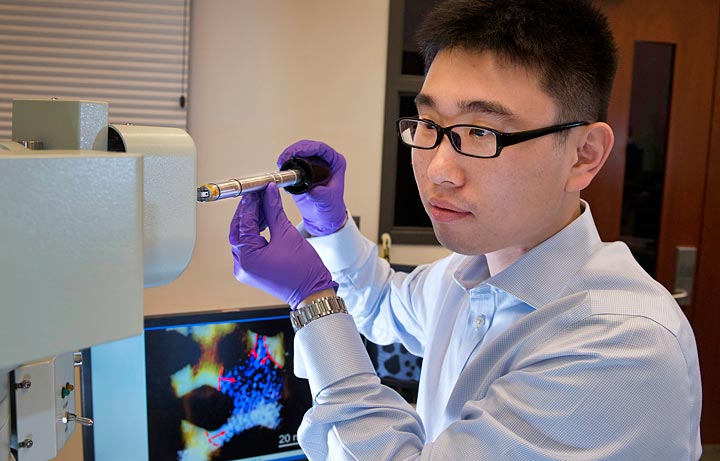
Materials scientist Huolin Xin in Brookhaven Lab's Center for Functional Nanomaterials.
Batteries do not age gracefully. The lithium ions that power portable electronics cause lingering structural damage with each cycle of charge and discharge, making devices from smartphones to tablets tick toward zero faster and faster over time. To stop or slow this steady degradation, scientists must track and tweak the imperfect chemistry of lithium-ion batteries with nanoscale precision.
“We discovered surprising and never-before-seen evolution and degradation patterns in two key battery materials,” said Huolin Xin, a materials scientist at Brookhaven Lab’sCenter for Functional Nanomaterials(CFN) and coauthor on both studies. “Contrary to large-scale observation, the lithium-ion reactions actually erode the materials non-uniformly, seizing upon intrinsic vulnerabilities in atomic structure in the same way that rust creeps unevenly across stainless steel.”In two recent Nature Communicationspapers, scientists from several U.S. Department of Energy national laboratories—Lawrence Berkeley, Brookhaven, SLAC, and the National Renewable Energy Laboratory—collaborated to map these crucial billionths-of-a-meter dynamics and lay the foundation for better batteries.
Scientists used electron tomography techniques to create this 3D animation of the nickel-oxide nanosheet transformations during the lithium-ion battery charging process.
Xin used world-leading electron microscopy techniques in both studies to directly visualize the nanoscale chemical transformations of battery components during each step of the charge-discharge process. In an elegant and ingenious setup, the collaborations separately explored a nickel-oxide anode and a lithium-nickel-manganese-cobalt-oxide cathode—both notable for high capacity and cyclability—by placing samples inside common coin-cell batteries running under different voltages.
“Armed with a precise map of the materials’ erosion, we can plan new ways to break the patterns and improve performance,” Xin said.
In these experiments, lithium ions traveled through an electrolyte solution, moving into an anode when charging and a cathode when discharging. The processes were regulated by electrons in the electrical circuit, but the ions’ journeys—and the battery structures—subtly changed each time.
Chinks in Nano-Armor
For the nickel-oxide anode, researchers submerged the batteries in a liquid organic electrolyte and closely controlled the charging rates. They stopped at predetermined intervals to extract and analyze the anode. Xin and his collaborators rotated 20-nanometer-thick sheets of the post-reaction material inside a carefully calibrated transmission electron microscope (TEM) grid at CFN to catch the contours from every angle—a process called electron tomography.
In the experimental coin cell setup, a carbon supported transmission electron microscopy (TEM) grid loaded with a small amount of the nickel-oxide material was pressed against the bulk anode and submerged in the same electrolyte environment.
To see the way the lithium-ions reacted with the nickel oxide, the scientists used a suite of custom-written software to digitally reconstruct the three-dimensional nanostructures with single-nanometer resolution. Surprisingly, the reactions sprang up at isolated spatial points rather than sweeping evenly across the surface.
“Consider the way snowflakes only form around tiny particles or bits of dirt in the air,” Xin said. “Without an irregularity to glom onto, the crystals cannot take shape. Our nickel oxide anode only transforms into metallic nickel through nanoscale inhomogeneities or defects in the surface structure, a bit like chinks in the anode’s armor.”
The electron microscopy provided a crucial piece of the larger puzzle assembled in concert with Berkeley Lab materials scientists and soft x-ray spectroscopy experiments conducted at SLAC’s Stanford Synchrotron Radiation Lightsource (SSRL). The combined data covered the reactions on the nano-, meso-, and microscales.
Rock-Salt Buildups
In the other study, scientists sought the voltage sweet-spot for the high-performing lithium-nickel-manganese-cobalt-oxide (NMC) cathode: How much power can be stored, at what intensity, and across how many cycles?
The answers hinged on intrinsic material qualities and the structural degradation caused by cycles at 4.7 volts and 4.3 volts, as measured against a lithium metal standard.
As revealed through another series of coin-cell battery tests, 4.7 volts caused rapid decomposition of the electrolytes and poor cycling—the higher power comes at a price. A 4.3-volt battery, however, offered a much longer cycling lifetime at the cost of lower storage and more frequent recharges.
In both cases, the chemical evolution exhibited sprawling surface asymmetries, though not without profound patterns.
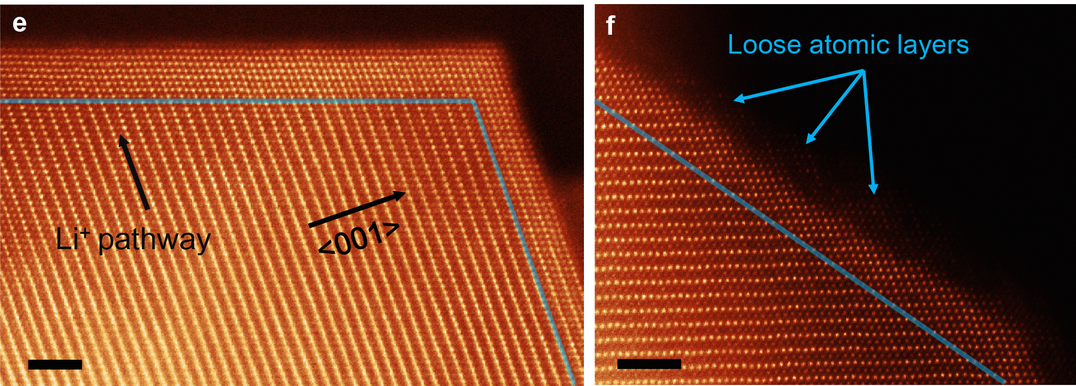
Each orange dot in these scanning transmission electron microscopy (STEM) images represents one atomic column in the NMC cathode. The scientists found that the lithium ions tended to travel along the vertical channels between atomic layers. After one full charge/discharge cycle, the surface layers (the edge beyond the blue line) exhibited the atomic disordering that ultimately diminishes battery performance.
“As the lithium ions race through the reaction layers, they cause clumping crystallization—a kind of rock-salt matrix builds up over time and begins limiting performance,” Xin said. “We found that these structures tended to form along the lithium-ion reaction channels, which we directly visualized under the TEM. The effect was even more pronounced at higher voltages, explaining the more rapid deterioration.”
Identifying this crystal-laden reaction pathways hints at a way forward in battery design.
“It may be possible to use atomic deposition to coat the NMC cathodes with elements that resist crystallization, creating nanoscale boundaries within the micron-sized powders needed at the cutting-edge of industry,” Xin said. “In fact, Berkeley Lab battery experts Marca Doeff and Feng Lin are working on that now.”
Shirley Meng, a professor at UC San Diego’s Department of NanoEngineering, added, “This beautiful study combines several complementary tools that probe both the bulk and surface of the NMC layered oxide—one of the most promising cathode materials for high-voltage operation that enables higher energy density in lithium-ion batteries. The meaningful insights provided by this study will significantly impact the optimization strategies for this type of cathode material.”
The TEM measurements revealed the atomic structures while electron energy loss spectroscopy helped pinpoint the chemical evolution—both carried out at the CFN. Further crucial research was conducted at SLAC’s SSRL and Berkeley Lab’s National Center for Materials Synthesis, Electrochemistry, and Electron Microscopy, with computational support from the National Energy Research Supercomputer Center and the Extreme Science and Engineering Discovery Environment.
Toward Real-Time, Real-World Analyses
“The chemical reactions involved in these batteries are startlingly complex, and we need even more advanced methods of interrogation,” Xin said. “My CFN colleagues are developing ways to watch the reactions in real-time rather than the stop-and-go approach we used in these studies.”
These in operando microscopy techniques, led in part by Brookhaven Lab materials scientists Dong Su, Feng Wang, and Eric Stach, will image reactions as they unfold in liquid environments. Custom-designed electrochemical contacts and liquid flow holders will usher in unprecedented insights.
Research at Brookhaven Lab’s CFN and SLAC’s SSRL—both DOE user facilities—was supported by DOE’s Office of Science. The NMC work was also supported through the Batteries for Advanced Transportation Technologies (BATT) program funded by DOE’s Office of Energy Efficiency and Renewable Energy and led by Berkeley Lab.
DOE's Office of Science is the single largest supporter of basic research in the physical sciences in the United States, and is working to address some of the most pressing challenges of our time. For more information, please visit science.energy.gov.