Showing posts with label bacteria. Show all posts

Friday, December 18, 2015

Nanotech weapon against chronic bacterial infections in hospitals


One of the scourges of infections in hospitals -- biofilms formed by bacteria that stick to each other on living tissue and medical instruments, making them harder to remove -- can be tricked into dispersing with the targeted application of nanoparticles and heat, researchers have found.

The University of New South Wales study, jointly led by Associate Professor Cyrille Boyer of the School of Chemical Engineering and deputy director of Australian Centre for NanoMedicine, appears in today's issue of Nature's open access journal Scientific Reports.

"Chronic biofilm-based infections are often extremely resistant to antibiotics and many other conventional antimicrobial agents, and have a high capacity to evade the body's immune system," said Boyer. "Our study points to a pathway for the non-toxic dispersal of biofilms in infected tissue, while also greatly improving the effect of antibiotic therapies." 

Biofilms have been linked to 80% of infections, forming on living tissues (eg. respiratory, gastrointestinal and urinary tracts, oral cavities, eyes, ears, wounds, heart and cervix) or dwelling in medical devices (eg. dialysis catheters, prosthetic implants and contact lenses).

The formation of biofilms is a growing and costly problem in hospitals, creating infections that are more difficult to treat -- leading to chronic inflammation, impaired wound healing, rapidly acquired antibiotic resistance and the spread of infectious embolisms in the bloodstream.

They also cause fouling and corrosion of wet surfaces, and the clogging of filtration membranes in sensitive equipment -- even posing a threat to public health by acting as reservoirs of pathogens in distribution systems for drinking water.

In general, bacteria have two life forms during growth and proliferation: planktonic, where bacteria exist as single, independent cells; or aggregated together in colonies as biofilms, where bacteria grow in a slime-like polymer matrix that protects them from the environment around them.

Acute infections mostly involve planktonic bacteria, which are usually treatable with antibiotics.

However, when bacteria have had enough time to form a biofilm -- within a human host or non-living material such as dialysis catheters -- an infection can often become untreatable and develop into a chronic state.

Although biofilms were first recognised in the 17th century, their importance was not realised until the 1990s, when it became clear that microbes exist in nature more often in colonies made up of lots of different microorganisms that adhere to surfaces through slime excreted by their inhabitants. Thus began a global race to understand biofilms, at a time when it was also realised they were responsible for the majority of chronic infections.

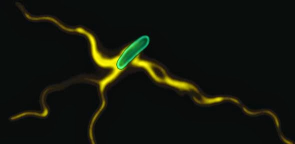
The discovery of how to dislodge biofilms by the UNSW team - jointly led by Dr Nicolas Barraud, formerly of UNSW and now at France's Institut Pasteur -- was made using the opportunistic human pathogen Pseudomonas aeruginosa. This is a model organism whose response to the technique the researchers believe will apply to most other bacteria.

When biofilms want to colonise a new site, they disperse into individual cells, reducing the protective action of the biofilm. It is this process the UNSW team sought to trigger, making the bacteria again susceptible to antimicrobial agents.

The UNSW team found that by injecting iron oxide nanoparticles into the biofilms, and using an applied magnetic field to heat them -- which induces local hyperthermia through raising the temperature by 5°C or more - the biofilms were triggered into dispersing.

They achieved this using iron oxide nanoparticles coated with polymers that help stabilise and maintain the nanoparticles in a dispersed state, making them an ideal non-toxic tool for treating biofilm infections.

"The use of these polymer-coated iron oxide nanoparticles to disperse biofilms may have broad applications across a range of clinical and industrial settings," said Boyer, who in October was named Physical Scientist of the Year in Australia's Prime Minister's Prizes for Science.
"Once dispersed, the bacteria are easier to deal with - creating the potential to remove recalcitrant, antimicrobial-tolerant biofilm infections."


Tuesday, February 17, 2015

Bacterial Armor Holds Clues for Self-Assembling Nanostructures


Many bacteria and archaea encase themselves within a self-assembling protective shell of S-layer proteins, like chainmail armor. The process is a model for the self-assembly of 2D and 3D organic and inorganic nanostructures.
Imagine thousands of copies of a single protein organizing into a coat of chainmail armor that protects the wearer from harsh and ever-changing environmental conditions. That is the case for many microorganisms. In a new study, researchers with the U.S. Department of Energy (DOE)’s Lawrence Berkeley National Laboratory (Berkeley Lab) have uncovered key details in this natural process that can be used for the self-assembly of nanomaterials into complex two- and three-dimensional structures.
Caroline Ajo-Franklin, a chemist and synthetic biologist at Berkeley Lab’s Molecular Foundry, led this study in which high-throughput light scattering measurements were used to investigate the self-assembly of 2D nanosheets from a common bacterial surface layer (S-layer) protein. This protein, called “SbpA,” forms the protective armor for Lysinibacillus sphaericus, a soil bacterium used as a toxin to control mosquitoes. Their investigation revealed that calcium ions play a key role in how this armor assembles. Two key roles actually.
“Calcium ions not only trigger the folding of the protein into the correct shape for nanosheet formation, but also serve to bind the nanosheets together,” Ajo-Franklin says. “By establishing and using light scattering as a proxy for SbpA nanosheet formation, we were able to determine how varying the concentrations of calcium ions and SbpA affects the size and shape of the S-layer armor.”
Caroline Ajo-Franklin, Steve Whitelam and Behzad Rad led a team at Berkeley Lab’s Molecular Foundry that uncovered key details by which bacterial proteins self-assemble into a protective armor coating. (Photo by Roy Kaltschmidt)
Details on this study have been published in the journal ACS Nano in a paper titled “Ion-Specific Control of the Self-Assembly Dynamics of a Nanostructured Protein Lattice.” Ajo-Franklin is the corresponding author. Co-authors are Behzad Rad, Thomas Haxton, Albert Shon, Seong-Ho Shin and Stephen Whitelam.
In the microbial world of bacteria and archaea, external threats abound. Their surrounding environment can transition from extreme heat to extreme cold, or from highly acidic to highly basic. Predators are everywhere. To protect themselves, many bacteria and archaea encase themselves within a shell of S-layer proteins. While scientists have known about this protective coating for many years, how it forms has been a mystery.
Ajo-Franklin and her colleagues have been exploring self-assembling proteins as a potential means of creating nanostructures with complex structure and function.
“At the Molecular Foundry, we’ve gotten really good at making nanomaterials into different shapes but we are still learning how to assemble these materials into organized structures,” she says. “S-layer proteins are abundant biological proteins known to self-assemble into 2D crystalline nanosheets with lattice symmetries and pore sizes that are about the same dimensions as quantum dots and nanotubes. This makes them a compelling model system for the creation of nanostructured arrays of organic and inorganic materials in a bottom-up fashion.”
The binding of calcium ions to SbpA proteins starts the process by which the SbpA self-assembles into nanosheets. Ca2+ binds to SbpA with an affinity of 67 μM.
The binding of calcium ions to SbpA proteins starts the process
by which the SbpA self-assembles into nanosheets. Ca2+ binds to
SbpA with an affinity of 67 μM.
In this latest study, light-scattering measurements were used to map out diagrams that revealed the relative yield of self-assembled nanosheets over a wide range of concentrations of SbpA and calcium ions. In addition, the effects of substituting manganese or barium ions for calcium ions were examined to distinguish between a chemically specific and generic divalent cation role for the calcium ions. Behzad Rad, the lead author of the ACS Nano paper, and co-workers followed light-scattering by light in the visible spectrum. They then correlated the signal to nanosheet formation by using electron microscopy and Small Angle X-ray Scattering (SAXS), a technology that can provide information on molecular assemblies in just about any type of solution. The SAXS measurements were obtained at the “SIBYLS beamline (12.3.1) of Berkeley Lab’s Advanced Light Source.

“We learned that only calcium ions trigger the SbpA self-assembly process and that the concentrations of calcium ions inside the cell are too low for nanosheets to form, which is a good thing for the bacterium,” says Rad. “We also found that the time evolution of the light scattering traces is consistent with the irreversible growth of sheets from a negligibly small nucleus. As soon as five calcium ions bind to a SbpA protein, the process starts and the crystal grows really fast. The small nucleus is what makes our light-scattering technique work.”
Ajo-Franklin, Rad and their co-authors believe their light-scattering technique is applicable to any type of protein that self-assembles into 2D nanosheets, and can be used to monitor growth from the nanometer to the micrometer scales.
Given the rugged nature of the S-layer proteins and their adhesive quality – bacteria use their S-layer armor to attach themselves to their surroundings – there are many intriguing applications awaiting further study.
“One project we’re exploring is using SbpA proteins to make adhesive nanostructures that could be used to remove metals and other contaminants from water,” Ajo-Franklin says. “Now that we have such a good handle on how SbpA proteins self-assemble, we’d like to start mixing and matching them with other molecules to create new and useful structures.”
http://newscenter.lbl.gov/2015/02/11/bacterial-armor/

Monday, March 10, 2014

Scientists combine bacteria with liquid crystals


When swimming around, bacteria aren’t good with the “pool rules.”  In small quantities, they’ll follow the lanes, but put enough together and they’ll begin to create their own flow.
In a collaboration between the U.S. Department of Energy’s Argonne National Laboratory and the Liquid Crystal Institute at Kent State University, researchers combined bacteria with liquid crystals and observed how the bacteria swam around and interacted with the medium, forming “living liquid crystals” that may have interesting applications for new material design and fabrication.
Liquid crystals are intermediate materials that exhibit some of the behaviors of a liquid and some of those of a crystalline solid. For example, a liquid crystal has a liquid-like flow, but on the molecular level it may appear more crystalline.
When they are dissolved in water, the molecules that make up a liquid crystal tend to form long rod-like structures that prefer to organize along one dimension, called a director. The director is symmetrical -- it does not have poles along its axis.
Using a solution known as “Terrific Broth” – so known for its usefulness as a bacterial growth medium – the researchers cultured a colony ofBacillus subtilis bacteria and transferred it to a liquid crystal.
When the researchers looked at the bacteria in the liquid crystal using a powerful optical microscope, they saw that they moved in a surprising way. Each B. subtilis bacterium has a long, thin tail called a flagellum, which the bacterium spins like a screw to propel itself. Although the bacteria originally orient themselves parallel to the director, the spinning of the flagella as the bacteria swim locally changes the director’s alignment into a wave-like pattern.
As the researchers increased the density of bacteria in the liquid crystal solution, they saw a second surprising effect. Because of the collective effects, the swimming bacteria began to create “stripes” of different director orientations within the liquid crystal. These stripes could then be erased by depriving the bacteria of oxygen.
“The system is extremely sensitive,” said Argonne materials scientist Andrey Sokolov, one of the authors of the study. “There is always an interplay between the bacterial forces and liquid crystal forces, and the behavior of the material depends on which one wins in the end.”
According to Argonne materials scientist Igor Aronson, the study’s lead author, it only takes a bacterial concentration of 0.2% for the bacteria’s collective swimming to begin to overtake the natural order of the liquid crystal.
The researchers hope that eventually studies on living liquid crystals as well as other similar “living materials” will pay dividends for a number of different applications. “Our principal goal is understanding active materials,” Aronson said. “We want to see how we can design new ways to consume energy from the environment and use it for self-assembly and self-repair.”
The scientists also noted that their method may have other implications beyond the living liquid crystals themselves.  Because the wake created by the oscillations of the flagella in the liquid crystal is so much larger than the nanometer-wide flagella themselves, the researchers were able to observe the action of the flagella with an optical microscope. “The flagella are only a few tens of nanometers in diameter, and in the past we’ve had to use electron microscopes in order to see it,” Aronson said.
“This could have bigger implications in terms of ways to visualize other nanoscale objects,” Sokolov added.
Funding for the study was provided by DOE’s Office of Science and the National Science Foundation, and results appeared as a paper titled "Living liquid crystals" in the Proceedings of the National Academy of Sciences.
Argonne National Laboratory seeks solutions to pressing national problems in science and technology. The nation's first national laboratory, Argonne conducts leading-edge basic and applied scientific research in virtually every scientific discipline. Argonne researchers work closely with researchers from hundreds of companies, universities, and federal, state and municipal agencies to help them solve their specific problems, advance America's scientific leadership and prepare the nation for a better future. With employees from more than 60 nations, Argonne is managed by UChicago Argonne, LLC for the U.S. Department of Energy's Office of Science.
The DOE Office of Science is the single largest supporter of basic research in the physical sciences in the United States and is working to address some of the most pressing challenges of our time. For more information, please visit http://science.energy.gov.
Source: http://www.anl.gov/articles/it-s-alive-scientists-combine-bacteria-liquid-crystals

Thursday, February 13, 2014

Computer Chips that Listen to Bacteria

In a study published today in Nature Communications, a research team led by Ken Shepard, professor of electrical engineering and biomedical engineering at Columbia Engineering, and Lars Dietrich, assistant professor of biological sciences at Columbia University, has demonstrated that integrated circuit technology, the basis of modern computers and communications devices, can be used for a most unusual application—the study of signaling in bacterial colonies. They have developed a chip based on complementary metal-oxide-semiconductor (CMOS) technology that enables them to electrochemically image the signaling molecules from these colonies spatially and temporally. In effect, they have developed chips that “listen” to bacteria.
“This is an exciting new application for CMOS technology that will provide new insights into how biofilms form,” says Shepard. “Disrupting biofilm formation has important implications in public health in reducing infection rates.”
The researchers, who include PhD students Daniel Bellin (electrical engineering) and Hassan Sakhtah (biology), say that this is the first time integrated circuits have been used for such an application—imaging small molecules electrochemically in a multicellular structure. While optical microscopy techniques remain paramount for studying biological systems (using photons allows for relatively non-invasive interaction to the biological system being studied), they cannot directly detect critical components of physiology, such as primary metabolism and signaling factors.
The team thought there might be a better way to directly detect small molecules through techniques that employ direct transduction to electrons, without using photos as an intermediary. They made an integrated circuit, a chip that, Shepard says, is an ‘active’ glass slide, a slide that not only forms a solid-support for the bacterial colony but also ‘listens’ to the bacteria as they talk to each other.”
“This is a big step forward,” Dietrich continues. “We describe using this chip to ‘listen in’ on conversations taking place in biofilms, but we are also proposing to use it to interrupt these conversations and thereby disrupt the biofilm. In addition to the pure science implications of these studies, a potential application of this would be to integrate such chips into medical devices that are common sites of biofilm formation, such as catheters, and then use the chips to limit bacterial colonization.”Cells, Dietrich explains, mediate their physiological activities using secreted molecules. The team looked specifically at phenazines, which are secreted metabolites that control gene expression. Their study found that the bacterial colonies produced a phenazine gradient that, they say, is likely to be of physiological significance and contribute to colony morphogenesis.
The next step for the team will be to develop a larger chip that will enable larger colonies to be imaged at higher spatial and temporal resolutions.
“This represents a new and exciting way in which solid-state electronics can be used to study biological systems,” Shepard adds. “This is one of the many emerging ways integrated circuit technology is having impact in biotechnology and the life sciences.”
The study was supported by the National Institutes of Health and the National Science 

Source: http://engineering.columbia.edu/chips-listen-bacteria-0

Monday, January 27, 2014

Novel biological mechanism relays electrons in proteins in mineral-breathing bacteria

Researchers at Pacific Northwest National Laboratory and University College London used EMSL and other computational resources to simulate how electrical current moves through molecular wires, called hemes, in certain bacteria. The findings are central to understanding bacterial ground chemistry and the use of bacteria for microbial fuel cells, batteries and turning waste into electricity.
This is the first time scientists have seen this evolutionary design principle for electron transport, the researchers reported inProceedings of the National Academy of Sciences Early Edition Online.

Researchers simulating how certain bacteria run electrical current through tiny molecular wires have discovered a secret Nature uses for electron travel. The results are key to understanding how the bacteria do chemistry in the ground, and will help researchers use them in microbial fuel cells, batteries, or for turning waste into electricity.
Within the bacteria's protein-based wire, molecular groups called hemes communicate with each other to allow electrons to hop along the chain like stepping stones. The researchers found that evolution has set the protein up so that, generally, when the electron's drive to hop is high, the heme stepping stones are less tightly connected, like being farther apart; when the drive to hop is low, the hemes are more closely connected, like being closer together. The outcome is an even electron flow along the wire.
This is the first time scientists have seen this evolutionary design principle for electron transport, the researchers reported Jan. 2 inProceedings of the National Academy of Sciences Early Edition Online.
"We were perplexed at how weak the thermodynamic driving force was between some of these hemes," said geochemist Kevin Rosso of the Department of Energy's Pacific Northwest National Laboratory. "But it turns out those pairs of hemes are essentially hugging each other. When the driving force is strong between hemes, they are only shaking hands. We've never seen this compensation scheme before, but it seems that the purpose is to allow the protein to transfer electrons with a steady flow along heme wires."
Living Wires
Certain bacteria breathe using metal like people use oxygen. In the process, these bacteria steal electrons from carbon and ultimately transfer the electrons to metals or minerals in the ground. They do this by conducting electricity along molecular wires built into proteins, moving internal electrons to the outside of their cells. Researchers hope to use these bacteria in little biologic batteries or fuel cells.
But a living wire is not the same as those that make up our powerlines. Electrons in powerlines hurtle down the wire, moving smoothly from metal atom to metal atom. Electrons traveling in a living wire must get from one complex heme group to the next. The hemes are situated within a protein, and not all hemes are made the same.
Some hemes hold onto electrons tightly and others let electrons slip away easily. Depending on how the hemes are lined up, this can create energetic hills that electrons have a hard time climbing over, or energetic valleys that electrons easily march across.
Some hemes, such as those that carry oxygen in people's red blood cells, are well-studied. The hemes and proteins creating a current in bacteria, though, have only been coming to light within the last few years. Recently, researchers figured out what a particular protein — MtrF — that makes up a molecular wire looks like, but that information alone is not enough to determine how the electrons traverse the chain of internal heme groups.
So, armed with the structure of the protein, Rosso and colleagues Jochen Blumberger and Marian Breuer from the University College London used high-powered computers to simulate the positions and movement of the hemes in MtrF and how they transfer electrons between themselves.
Electron Crossroads
Using resources at both the UK's High Performance Computing Facility and EMSL, the Environmental Molecular Sciences Laboratory at PNNL, the team first modeled the average position of the 10 hemes within MtrF. Eight of the hemes run down the center of the protein. The remaining two hemes branch off the main eight, creating a four-heme road that crosses the middle of the protein.
Because hemes have to pass electrons to each other, the team examined them in pairs. The team found that MtrF arranges its heme pairs in one of three ways: perpendicular to each other, side-by-side, or stacked on top of each other. Each arrangement positions the hemes at different distances from and orientations to each other.
Then the team gauged how urgently an electron wants to get from one heme to the next by determining the theoretical "Gibbs free energy" between the pairs. This value is an indicator of the driving force of the electrons.
The team found that instead of a smooth ride through the protein, electrons lurch through hemes: Sometimes the driving force makes the electrons march across a valley and the electrons move quickly. In other pairs the electrons face a hill, and electron travel gets delayed.
Mapping how tightly hemes couple to each other along with the driving force values, the team found that hemes were less tightly coupled when electrons enjoyed traipsing across a valley and more tightly coupled when electrons had to slog uphill.
"The computer simulations allowed us to break the wire down into how each step is possible and how fast each step is. Then we saw that the protein arranges its hemes in weak and strong couplings to compensate for the energetic hills and valleys," said Rosso. "This is one way to make the electron hops consistent to efficiently get them where they need to go."
This compensation scheme led the team to wonder why the hills and valleys are there in the first place.
"We think the variation in driving force between the hills and the valleys helps the protein interact with other components in the environment," said Rosso. The tops of the hills could be exit points to higher energy electron acceptors in the environment, such as molecules that shuttle electrons elsewhere.
Scientists don't yet know how multiple heme proteins — including others beyond MtrF — work in concert to make these molecular wires connect end-to-end, but the results give hints as to which hemes are possible entry and exit points in MtrF. So the results also give clues to how multiple proteins might be connected.
This work was supported by the Department of Energy Office of Science. Support for use of the UK's High Performance Computing Facility was provided by the UK's Engineering and Physical Sciences Research Council. Additional support was provided by the Royal Society.

Souce: http://www.pnnl.gov/news/release.aspx?id=1032

Monday, January 6, 2014

Tiny acts of microbe justice help reveal how nature fights freeloaders

Princeton University researchers discovered that the bacteria Vibrio cholerae 
keeps food generated by the community's productive members away 
from those of their kind that attempt to live on others' leftovers. The bacteria use 
two mechanisms that are likely common among bacteria. In some instances,
the natural flow of fluids over the surface of bacterial communities can wash away 
excess food before the freeloaders can indulge. In microscope images, 
shiftless V. cholerae (red) were in abundance under conditions of no fluid flow 
(left image). When the bacteria were grown in an environment with fluid 
flow — similar to that found in nature — cooperative V. cholerae (yellow) 
won out (right image). 
(Image courtesy of Carey Nadell, Department of Molecular Biology)
The idea of everyone in a community pitching in is so universal that even bacteria have a system to prevent the layabouts of their kind from enjoying the fruit of others' hard work, Princeton University researchers have discovered.
Groups of the bacteria Vibrio cholerae deny loafers their unjust desserts by keeping the food generated by the community's productive members away from V. cholerae that attempt to live on others' leftover nutrients, the researchers report in the journal Current Biology. The researchers found that individual bacteria produce a thick coating around themselves to prevent nutrients from drifting over to the undeserving. Alternatively, the natural flow of fluids over the surface of bacterial communities can wash away excess food before the freeloaders can indulge.
Likely common among bacteria, this act of microscopic justice not only ensures the survival of the group's most industrious members, but also could be used for agriculture, fuel production and the treatment of bacterial infections such as cholera, explained first author Knut Drescher, a postdoctoral research fellow in the lab of senior author Bonnie Bassler, the Squibb Professor in Molecular Biology and department chair.
By encouraging this action, scientists could increase the efficiency of any process that relies on bacteria to break down organic materials, such as plant materials into biofuels, or cellulose into paper products, Drescher said. For treating a disease, the mechanism could be counteracted to effectively starve the more productive bacteria and weaken the infection.
"We could use our discovery to develop strategies that encourage the proliferation of microbes that digest dead organic material into useful products," Drescher said. "Such an approach will be useful for optimizing nutrient recycling for agriculture, bioremediation, industrial cleanup, or making products for industry or medicine."
The Princeton findings also provide insight into how all microbes potentially preserve themselves by imposing fairness and resolving the "public goods dilemma," in which a group must work together while also avoiding exploitation by their self-serving individuals, said co-lead author Carey Nadell, a postdoctoral research associate in Bassler's lab.
"The public goods dilemma is a central problem in the history of life on Earth, during which single cells have emerged as collectives of genes, multicellular organisms have emerged as collectives of cells, and societies have emerged as collectives of multicellular organisms," Nadell said.
"At each of these transitions in complexity there has been — and remains — the threat of exploitation by single members pursuing their own interests at the expense of the collective as a whole," Nadell said. "Clarifying how exploitation can be averted is therefore critical to understanding how life has taken the various forms that exist today."
Bassler figure 3
All bacteria frequently live in dense communities called biofilms. They secrete enzymes that break down solid organic carbon- and nitrogen-containing molecules and feast on the components within. But not every individual bacterium will produce enzymes — some will simply feed on what their neighbors produce. The Princeton researchers found that individual bacteria also will produce a thick coating around themselves to prevent nutrients from drifting over to the undeserving. In the thicker biofilms near the top of this microscope image, productive V. cholerae (yellow) overtook exploitive V. cholerae (red). The darker communities indicate thinner biofilms and a proliferation of bacteria that will live off the work of others. (Image courtesy of Carey Nadell, Department of Molecular Biology)
Like all bacteria, V. cholerae — strains of which can cause cholera — frequently lives in dense communities called biofilms. Also like other bacteria, V. cholerae secretes enzymes that break down the solid organic carbon- and nitrogen-containing molecules of which living things are composed so that the bacterium can feast on the components within. But not every individual bacterium will produce enzymes — some will simply feed on what their organic-compound digesting neighbors produce. The researchers found two mechanisms by which this leeching is halted.
The vigilance of V. cholerae and other bacteria may also carry a larger benefit. The nitrogen and carbon that make up most of the planet's breathable air largely come from the digestion of organic materials by bacteria.
The researchers studied V. cholerae as it feasted on its preferred victual, chitin, a sugar-based molecule and the central element of many marine cells, exoskeletons and other appendages. The researchers write that sea animals alone shed an estimated 110 billion tons of chitin each year — yet hardly any of it makes it to the ocean floor. Instead, the detritus is consumed by V. cholerae and other marine bacteria with its elements being recycled into the biosphere.
"If V. cholerae's system of extracellular digestion were compromised by exploitation," Nadell said, "the world's supply of carbon and nitrogen would become sequestered on a rapid geological timescale."
Drescher, Nadell and Bassler worked with Howard Stone, the Donald R. Dixon '69 and Elizabeth W. Dixon Professor in Mechanical and Aerospace Engineering, and Ned Wingreen, the Howard A. Prior Professor in the Life Sciences and the Lewis-Sigler Institute for Integrative Genomics.
The paper, "Solutions to the Public Goods Dilemma in Bacterial Biofilms," was published Jan. 4 in the journal Current Biology. The work was supported by grants from the Howard Hughes Medical Institute, the National Institutes of Health (grant 5R01GM065859), the National Science Foundation (grants MCB-0343821 and MCB- 1119232), and the Human Frontier Science Program.

Monday, November 18, 2013

Building ‘nanomachines’ in biological outer space


New research reveals how bacteria construct tiny flagella ‘nanomachines’ outside the cell

Cambridge scientists have uncovered the mechanism by which bacteria build their surface propellers (flagella) – the long extensions that allow them to swim towards food and away from danger. The results, published this week in the journal Nature, demonstrate how the mechanism is powered by the subunits themselves as they link in a chain that is pulled to the flagellum tip.

Previously, scientists thought that the building blocks for flagella were either pushed or diffused from the flagellum base through a central channel in the structure to assemble at the flagellum tip, which is located far outside the cell. However, these theories are incompatible with recent research showing that flagella grow at a constant rate. The completely new and unexpected chain mechanism, in which subunits linked in a chain ‘pull themselves’ through the flagellum, transforms understanding of how flagellum assembly is energised.

The research was led by Dr Gillian Fraser and Professor Colin Hughes in the University’s Department of Pathology and was funded by the Wellcome Trust.

Dr Lewis Evans, who carried out the research, remarked: “It’s exciting how economical bacteria are, able to harness the thermal free energy from unfolded subunits and convert it into a coherent directed transport. More broadly, it’s fascinating to imagine the implications for how heat energy (normally considered as ‘lost’) might be harnessed to drive processes even outside living cells.”

As each flagellum ‘nanomachine’ is assembled, thousands of subunit ‘building blocks’ are made in the cell and are then unfolded and exported across the cell membrane. Like other processes inside cells, this initial export phase consumes chemical energy. However, when subunits pass out of the cell into the narrow channel at the center of the growing flagellum, there is no conventional energy source and they must somehow find the energy to reach the tip.

The team has shown that at the base of the flagellum, subunits connect by head-to-tail linkage into a long chain. Together with Professor Eugene Terentjev, at the Cavendish Laboratory, they showed that the chain is pulled through the entire length of the flagellum channel by the entropic force of the unfolded subunits themselves. This produces tension in the subunit chain, which increases as each subunit refolds and incorporates into the tip of the growing structure. This pulling force automatically adjusts with increasing flagellum length, providing a constant rate of subunit delivery to the assembly site at the tip.

Professor Terentjev noted that this breakthrough can be applied to other fields. “Understanding how polymers move through channels is a fundamental physical problem. Gaining insight into this has potential applications in other disciplines, for instance in nanotechnology, specifically the building of new nanomaterials.”

This research has far-reaching implications, according to Fraser. “By understanding the base-level, fundamental biology of medically important bacteria and their construction of flagella and related toxin-injecting molecular syringes,” she commented, “we can start to develop new ways to counteract them.”

Dr Gillian Fraser is at Queens' College; Professor Colin Hughes is at Trinity College; Professor Eugene Terentjev is at Queens' College

Source: 
http://www.cam.ac.uk/research/news/building-nanomachines-in-biological-outer-space