Showing posts with label cell. Show all posts

Friday, May 30, 2014

Hitchhiking nanotubes show how cells stir themselves

Rice, Göttingen, VU researchers track single-molecule proteins in living cells 

Chemical engineers from Rice University and biophysicists from Georg-August Universität Göttingen in Germany and the VU University Amsterdam in the Netherlands have successfully tracked single molecules inside living cells with carbon nanotubes.
Through this new method, the researchers found that cells stir their interiors using the same motor proteins that serve in muscle contraction.
The study, which sheds new light on biological transport mechanisms in cells, appears this week in Science.
The team attached carbon nanotubes to transport molecules known as kinesin motors to visualize and track them as they moved through the cytoplasm of living cells.
“I am amazed how versatile carbon nanotubes are,” said co-author Matteo Pasquali, a Rice professor of chemical and biomolecular engineering and of chemistry. “We use them for a wide range of applications, from engineering conducting fibers to imaging in cells.”
Carbon nanotubes are hollow cylinders of pure carbon with one-atom-thick walls. They naturally fluoresce with near-infrared wavelengths when exposed to visible light, a property discovered at Rice by Professor Rick Smalley a decade ago and then leveraged by Rice Professor Bruce Weisman to image carbon nanotubes. When attached to a molecule, the hitchhiking nanotubes serve as tiny beacons that can be precisely tracked over long periods of time to investigate small, random motions inside cells.
“Any probe that can hitch the length and breadth of the cell, rough it, slum it, struggle against terrible odds, win through and still know where its protein is, is clearly a probe to be reckoned with,” said lead author Nikta Fakhri, paraphrasing “The Hitchhiker’s Guide to the Galaxy.” Fakhri, who earned her Rice doctorate in Pasquali’s lab in 2011, is currently a Human Frontier Science Program Fellow at Göttingen.
“In fact, the exceptional stability of these probes made it possible to observe intracellular motions from times as short as milliseconds to as long as hours,” she said.
For long-distance transport, such as along the long axons of nerve cells, cells usually employ motor proteins tied to lipid vesicles, the cell’s “cargo containers.” This process involves considerable logistics: Cargo needs to be packed, attached to the motors and sent off in the right direction.
“This research has helped uncover an additional, much simpler mechanism for transport within the cell interior,” said principal investigator Christoph Schmidt, a professor of physics at Göttingen. “Cells vigorously stir themselves, much in the way a chemist would accelerate a reaction by shaking a test tube. This will help them to move objects around in the highly crowded cellular environment.”
The researchers showed the same type of motor protein used for muscle contraction is responsible for stirring. They reached this conclusion after exposing the cells to drugs that suppressed these specific motor proteins. The tests showed that the stirring was suppressed as well.
The mechanical cytoskeleton of cells consists of networks of protein filaments, like actin. Within the cell, the motor protein myosin forms bundles that actively contract the actin network for short periods. The researchers found random pinching of the elastic actin network by many myosin bundles resulted in the global internal stirring of the cell. Both actin and myosin play a similar role in muscle contraction. 
The highly accurate measurements of internal fluctuations in the cells were explained in a theoretical model developed by VU co-author Fred MacKintosh, who used the elastic properties of the cytoskeleton and the force-generation characteristics of the motors.
“The new discovery not only promotes our understanding of cell dynamics, but also points to interesting possibilities in designing ‘active’ technical materials,” said Fakhri, who will soon join the Massachusetts Institute of Technology faculty as an assistant professor of physics. “Imagine a microscopic biomedical device that mixes tiny samples of blood with reagents to detect disease or smart filters that separate squishy from rigid materials.”
Co-authors of the study include graduate student Alok Wessel, technical assistant Charlotte Willms and research scientist Dieter Klopfenstein, all of the University of Göttingen.
The German Research Foundation, the Dutch Foundation for Fundamental Research on Matter, the Netherlands Organization for Scientific Research, the Welch Foundation, the National Science Foundation and the Human Frontier Science Program supported the research.
-30-

View a short movie of nanotube-tagged proteins moving via stirring inside cells:


A thin carbon nanotube is attached to a molecular motor (yellow) that moves along microtubule filaments (green) that form the transport network of cells. This transport occurs in the highly crowded environment of the cytoplasm that includes a network of actin filaments (red). The fluorescent nanotube serves as a beacon for both the transport along the microtubule, as well as the buffeting of the microtubule by the highly agitated surrounding cytoplasm. (Credit: M. Leunissen, Dutch Data Design)


- See more at: http://news.rice.edu/2014/05/29/hitchhiking-nanotubes-show-how-cells-stir-themselves/#sthash.cQhqzdGm.dpuf

Saturday, May 24, 2014

Repair protein’s DNA recognition motif

DNA replication – the process of copying the DNA each time a cell divides – must be completed accurately to avoid mutations that cause cancer and other diseases. The DNA damage response protein SMARCAL1 recognizes stalled replication “forks” and remodels the DNA to allow repair and restored replication. SMARCAL1 is essential to maintaining genome integrity during replication, but how it works is poorly understood.
Now, Brandt Eichman, Ph.D., and colleagues have determined the crystal structure of a region of SMARCAL1 (the HARP domain), which is fused to a motor domain. They used X-ray scattering to examine the conformation and assembly of the HARP domain in solution and found that the domain is conserved with DNA damage recognition domains from other DNA repair proteins. They showed that the HARP domain is a functional substitute for one of these regions and that mutations of predicted DNA-binding amino acids in the HARP domain reduced its ability to bind to replication forks and facilitate repair.
The studies, reported in the Proceedings of the National Academy of Sciences, uncovered a conserved recognition domain in DNA repair enzymes. This domain couples DNA recognition and remodeling and plays an important role in stabilizing replication forks and maintaining genome integrity.
The structure also illustrates the location of several SMARCAL1 mutations that cause Schimke immuno-osseous dysplasia (SIOD), a multi-system disorder characterized by growth defects, immune deficiencies and renal failure.
The findings are the latest in an ongoing collaboration between the teams of Eichman, associate professor of Biological Sciences and Biochemistry, David Cortez, Ph.D., professor of Biochemistry and Cancer Biology, and Walter Chazin, Ph.D., Chancellor’s Professor of Biochemistry and Chemistry. Together, the researchers aim to understand how DNA replication happens faithfully so that every cell ends up with exactly the same DNA – and without damaging mutations.
The research was supported by a pilot grant from the Vanderbilt Center in Molecular Toxicology and by National Institutes of Health grant CA136933.
http://news.vanderbilt.edu/2014/05/repair-proteins-dna-recognition-motif/

Tuesday, March 4, 2014

First Look at How Individual Staphylococcus Cells Adhere to Nanostructures

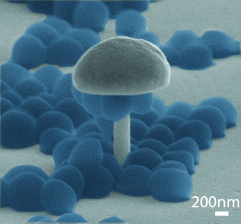
This scanning electron microscopy image reveals how Staphylococcus Aureus cells physically interact with a nanostructure. A bacterial cell (blue) is embedded inside the hollow nanopillar's hole and several cells cling to the nanopillar's curved walls. (Credit: Mofrad lab and the Nanomechanics Research Institute)


The bacterium Staphylococcus Aureus (S. aureus) is a common source of infections that occur after surgeries involving prosthetic joints and artificial heart valves. The grape-shaped microorganism adheres to medical equipment, and if it gets inside the body, it can cause a serious and even life-threatening illness called a Staph infection. The recent discovery of drug-resistant strains of S. aureus makes matters even worse.

A Staph infection can’t start unless Staphylococcus cells first cling to a surface, however, which is why scientists are hard at work exploring bacteria-resistant materials as a line of defense.
This research has now gone nanoscale, thanks to a team of researchers led by Berkeley Lab scientists. They investigated, for the first time, how individual S. aureus cells glom onto metallic nanostructures of various shapes and sizes that are not much bigger than the cells themselves.
They found that bacterial adhesion and survival rates vary depending on the nanostructure’s shape. Their work could lead to a more nuanced understanding of what makes a surface less inviting to bacteria.

Scanning electron microscopy image of bacterial cells (blue) suspended from the mushroom-shaped nanostructure's overhangs. (Credit: Mofrad lab and the Nanomechanics Research Institute)
“By understanding the preferences of bacteria during adhesion, medical implant devices can be fabricated to contain surface features immune to bacteria adhesion, without the requirement of any chemical modifications,” says Mohammad Mofrad, a faculty scientist in Berkeley Lab’s Physical Biosciences Division and a professor of Bioengineering and Mechanical Engineering at UC Berkeley.
Mofrad conducted the research with the Physical Biosciences Division’s Zeinab Jahed, the lead author of the study and a graduate student in Mofrad’s UC Berkeley Molecular Cell Biomechanics Laboratory, in collaboration with scientists from Canada’s University of Waterloo.
Their research was recently published online in the journal Biomaterials.
The scientists first used electron beam lithographic and electroplating techniques to fabricate nickel nanostructures of various shapes, including solid pillars, hollowed-out pillars, c-shaped pillars, and x-shaped columns. These features have outer diameters as small as 220 nanometers. They also created mushroom-shaped nanostructures with tiny stems and large overhangs.
They introduced S. aureus cells to these structures, gave the cells time to stick, and then rinsed the structures with deionized water to remove all but the most solidly bound bacteria.
Scanning electron microscopy revealed which shapes are the most effective at inhibiting bacterial adhesion. The scientists observed higher bacteria survival rates on the tubular-shaped pillars, where individual cells were partially embedded into the holes.

In contrast, pillars with no holes had the lowest survival rates.

The scientists also found that S. aureus cells can adhere to a wide range of surfaces. The cells not only adhere to horizontal surfaces, as expected, but to highly curved features, such as the sidewalls of pillars. The cells can also suspend from the overhangs of mushroom-shaped nanostructures.
“The bacteria seem to sense the nanotopography of the surface and form stronger adhesions on specific nanostructures,” says Jahed.

The research was supported by the Natural Sciences and Engineering Research Council of Canada and a National Science Foundation CAREER award.

###

Lawrence Berkeley National Laboratory addresses the world’s most urgent scientific challenges by advancing sustainable energy, protecting human health, creating new materials, and revealing the origin and fate of the universe. Founded in 1931, Berkeley Lab’s scientific expertise has been recognized with 13 Nobel prizes. The University of California manages Berkeley Lab for the U.S. Department of Energy’s Office of Science. For more, visit www.lbl.gov.

Source: http://newscenter.lbl.gov/feature-stories/2014/03/04/bacterial-adhesion/

Monday, November 25, 2013

Researchers Use Nanoscale ‘Patches’ to Sensitize Targeted Cell Receptors

Researchers from North Carolina State University and Duke University have developed nanoscale “patches” that can be used to sensitize targeted cell receptors, making them more responsive to signals that control cell activity. The finding holds promise for promoting healing and facilitating tissue engineering research.
The research takes advantage of the fact that cells in a living organism can communicate via physical contact. Specifically, when targeted receptors on the surface of a cell are triggered, the cell receives instructions to alter its behavior in some way. For example, the instructions may cause a stem cell to differentiate into a bone cell or a cartilage cell.
These receptors respond to specific ligands, or target molecules. And those ligands have to be present in certain concentrations in order to trigger the receptors. If there aren’t enough target ligands, the receptors won’t respond.
Now researchers have developed nanoscale patches that are embedded with tiny protein fragments called peptides. These peptides bond to a specific cell receptor, making it more sensitive to its target ligand – meaning that it takes fewer ligand molecules to trigger the receptor and its resulting behavior modification.
“This study shows that our concept can work, and there are a host of potential applications,” says Dr. Thom LaBean, an associate professor of materials science at NC State and senior author of a paper describing the work. “For example, if we identify the relevant peptides, we could create patches that sensitize cells to promote cartilage growth on one side of the patch and bone growth on the other side. This could be used to expedite healing or to enable tissue engineering of biomedical implants.”
“What’s important about this is that it allows us to be extremely precise in controlling cell behavior and gene expression,” says Ronnie Pedersen, a Ph.D. student at Duke University and lead author of the paper. “By controlling which peptides are on the patch, we can influence the cell’s activity. And by manipulating the placement of the patch, we can control where that activity takes place.”
The patch itself is made of DNA that researchers have programmed to self-assemble into flexible, two-dimensional sheets. The sheets themselves incorporate molecules called biotin and streptavidin which serve to hold and organize the peptides that are used to sensitize cell receptors.
“These peptides can bind with cell receptors and sensitize them, without blocking the interaction between the receptors and their target ligands,” Pedersen says. “That’s what makes this approach work.”
The paper, “Sensitization of Transforming Growth Factor-β Signaling by Multiple Peptides Patterned on DNA Nanostructures,” was published online Nov. 8 in the journal Biomacromolecules. The paper was co-authored by Dr. Elizabeth Loboa, associate professor of the joint biomedical engineering program at NC State and UNC-Chapel Hill. The work was supported by National Science Foundation grants DMS-CDI-0835794 and 1133427; National Institute of Biomedical Imaging and Bioengineering grant 1R03EB008790; and the Danish National Research Foundation.
-shipman-
Note to Editors: The study abstract follows.
“Sensitization of Transforming Growth Factor-β Signaling by Multiple Peptides Patterned on DNA Nanostructures”
Authors: Ronnie O. Pedersen, Duke University; Elizabeth G. Loboa, North Carolina State University and UNC-Chapel Hill; and Thomas LaBean, North Carolina State University
Published: online Nov. 8, Biomacromolecules
DOI: 10.1021/bm4011722
Abstract: We report sensitization of a cellular signaling pathway by addition of functionalized DNA nanostructures. Signaling by transforming growth factor β (TGFβ) has been shown to be dependent on receptor clustering. By patterning a DNA nanostructure with closely spaced peptides that bind to TGF? receptor, we observe increased sensitivity of NMuMG cells to TGFβ ligand. This is evidenced by translocation of secondary messenger proteins to the nucleus and stimulation of an inducible luciferase reporter at lower concentrations of TGFβ ligand. We believe this represents an important initial step toward realization of DNA as a self-assembling and biologically compatible material for use in tissue engineering and drug delivery.

Monday, November 18, 2013

Building ‘nanomachines’ in biological outer space


New research reveals how bacteria construct tiny flagella ‘nanomachines’ outside the cell

Cambridge scientists have uncovered the mechanism by which bacteria build their surface propellers (flagella) – the long extensions that allow them to swim towards food and away from danger. The results, published this week in the journal Nature, demonstrate how the mechanism is powered by the subunits themselves as they link in a chain that is pulled to the flagellum tip.

Previously, scientists thought that the building blocks for flagella were either pushed or diffused from the flagellum base through a central channel in the structure to assemble at the flagellum tip, which is located far outside the cell. However, these theories are incompatible with recent research showing that flagella grow at a constant rate. The completely new and unexpected chain mechanism, in which subunits linked in a chain ‘pull themselves’ through the flagellum, transforms understanding of how flagellum assembly is energised.

The research was led by Dr Gillian Fraser and Professor Colin Hughes in the University’s Department of Pathology and was funded by the Wellcome Trust.

Dr Lewis Evans, who carried out the research, remarked: “It’s exciting how economical bacteria are, able to harness the thermal free energy from unfolded subunits and convert it into a coherent directed transport. More broadly, it’s fascinating to imagine the implications for how heat energy (normally considered as ‘lost’) might be harnessed to drive processes even outside living cells.”

As each flagellum ‘nanomachine’ is assembled, thousands of subunit ‘building blocks’ are made in the cell and are then unfolded and exported across the cell membrane. Like other processes inside cells, this initial export phase consumes chemical energy. However, when subunits pass out of the cell into the narrow channel at the center of the growing flagellum, there is no conventional energy source and they must somehow find the energy to reach the tip.

The team has shown that at the base of the flagellum, subunits connect by head-to-tail linkage into a long chain. Together with Professor Eugene Terentjev, at the Cavendish Laboratory, they showed that the chain is pulled through the entire length of the flagellum channel by the entropic force of the unfolded subunits themselves. This produces tension in the subunit chain, which increases as each subunit refolds and incorporates into the tip of the growing structure. This pulling force automatically adjusts with increasing flagellum length, providing a constant rate of subunit delivery to the assembly site at the tip.

Professor Terentjev noted that this breakthrough can be applied to other fields. “Understanding how polymers move through channels is a fundamental physical problem. Gaining insight into this has potential applications in other disciplines, for instance in nanotechnology, specifically the building of new nanomaterials.”

This research has far-reaching implications, according to Fraser. “By understanding the base-level, fundamental biology of medically important bacteria and their construction of flagella and related toxin-injecting molecular syringes,” she commented, “we can start to develop new ways to counteract them.”

Dr Gillian Fraser is at Queens' College; Professor Colin Hughes is at Trinity College; Professor Eugene Terentjev is at Queens' College

Source: 
http://www.cam.ac.uk/research/news/building-nanomachines-in-biological-outer-space

Wednesday, October 23, 2013

Nanopore opens new cellular doorway for drug transport

A living cell is built with barriers to keep things out – and researchers are constantly trying to find ways to smuggle molecules in.‬ ‪Professor Giovanni Maglia (Biochemistry, Molecular and Structural Biology, KU Leuven) and his team have engineered a biological nanopore that acts as a selective revolving door through a cell's lipid membrane. The nanopore could potentially be used in gene therapy and targeted drug delivery.
‬‬‬‬‬‬‬
All living cells are enclosed by a lipid membrane that separates the interior of the cell from the outside environment. The influx of molecules through the cell membrane is tightly regulated by membrane proteins that act as specific doorways for the trafficking of ions and nutrients. Membrane proteins can also be used by cells as weapons. Such proteins attack a cell by making holes – nanopores – in 'enemy' cell membranes. Ions and molecules leak from the holes, eventually causing cell death.‬‬

‪Researchers are now trying to use nanopores to smuggle DNA or proteins across membranes. Once inside a cell, the DNA molecule could re-programme the cell for a particular action. Professor Maglia explains: "‪We are now able to engineer biological nanopores, but the difficult part is to precisely control the passage of molecules through the nanopores' doorways. We do not want the nanopore to let everything in. Rather, we want to limit entry to specific genetic information in specific cells." ‬‬‬‬‬‬‬‬‬‬‬‬‬‬

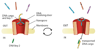
‪Professor Maglia and his team succeeded in engineering a nanopore that works like a revolving door for DNA molecules. "We have introduced a selective DNA revolving door atop of the nanopore. Specific DNA keys in solution hybridise to the DNA door and are transported across the nanopore. A second DNA key on the other side of the nanopore then releases the desired genetic information. A new cycle can then begin with another piece of DNA – as long as it has the correct key. In this way, the nanopore acts simultaneously as a filter and a conveyor belt." ‬‬‬‬‬‬‬

"In other words, we have engineered a selective transport system that can be used in the future to deliver medication into the cell. This could be of particular use in gene therapy, which involves introducing genetic material into degenerated cells in order to disable or re-programme them. It could also be used in targeted drug delivery, which involves administering medication directly into the cell. The possibilities are promising."‬‬

 
The researchers' findings were published in a recent edition of Nature Communications.