Showing posts with label protein. Show all posts

Friday, May 30, 2014

Hitchhiking nanotubes show how cells stir themselves

Rice, Göttingen, VU researchers track single-molecule proteins in living cells 

Chemical engineers from Rice University and biophysicists from Georg-August Universität Göttingen in Germany and the VU University Amsterdam in the Netherlands have successfully tracked single molecules inside living cells with carbon nanotubes.
Through this new method, the researchers found that cells stir their interiors using the same motor proteins that serve in muscle contraction.
The study, which sheds new light on biological transport mechanisms in cells, appears this week in Science.
The team attached carbon nanotubes to transport molecules known as kinesin motors to visualize and track them as they moved through the cytoplasm of living cells.
“I am amazed how versatile carbon nanotubes are,” said co-author Matteo Pasquali, a Rice professor of chemical and biomolecular engineering and of chemistry. “We use them for a wide range of applications, from engineering conducting fibers to imaging in cells.”
Carbon nanotubes are hollow cylinders of pure carbon with one-atom-thick walls. They naturally fluoresce with near-infrared wavelengths when exposed to visible light, a property discovered at Rice by Professor Rick Smalley a decade ago and then leveraged by Rice Professor Bruce Weisman to image carbon nanotubes. When attached to a molecule, the hitchhiking nanotubes serve as tiny beacons that can be precisely tracked over long periods of time to investigate small, random motions inside cells.
“Any probe that can hitch the length and breadth of the cell, rough it, slum it, struggle against terrible odds, win through and still know where its protein is, is clearly a probe to be reckoned with,” said lead author Nikta Fakhri, paraphrasing “The Hitchhiker’s Guide to the Galaxy.” Fakhri, who earned her Rice doctorate in Pasquali’s lab in 2011, is currently a Human Frontier Science Program Fellow at Göttingen.
“In fact, the exceptional stability of these probes made it possible to observe intracellular motions from times as short as milliseconds to as long as hours,” she said.
For long-distance transport, such as along the long axons of nerve cells, cells usually employ motor proteins tied to lipid vesicles, the cell’s “cargo containers.” This process involves considerable logistics: Cargo needs to be packed, attached to the motors and sent off in the right direction.
“This research has helped uncover an additional, much simpler mechanism for transport within the cell interior,” said principal investigator Christoph Schmidt, a professor of physics at Göttingen. “Cells vigorously stir themselves, much in the way a chemist would accelerate a reaction by shaking a test tube. This will help them to move objects around in the highly crowded cellular environment.”
The researchers showed the same type of motor protein used for muscle contraction is responsible for stirring. They reached this conclusion after exposing the cells to drugs that suppressed these specific motor proteins. The tests showed that the stirring was suppressed as well.
The mechanical cytoskeleton of cells consists of networks of protein filaments, like actin. Within the cell, the motor protein myosin forms bundles that actively contract the actin network for short periods. The researchers found random pinching of the elastic actin network by many myosin bundles resulted in the global internal stirring of the cell. Both actin and myosin play a similar role in muscle contraction. 
The highly accurate measurements of internal fluctuations in the cells were explained in a theoretical model developed by VU co-author Fred MacKintosh, who used the elastic properties of the cytoskeleton and the force-generation characteristics of the motors.
“The new discovery not only promotes our understanding of cell dynamics, but also points to interesting possibilities in designing ‘active’ technical materials,” said Fakhri, who will soon join the Massachusetts Institute of Technology faculty as an assistant professor of physics. “Imagine a microscopic biomedical device that mixes tiny samples of blood with reagents to detect disease or smart filters that separate squishy from rigid materials.”
Co-authors of the study include graduate student Alok Wessel, technical assistant Charlotte Willms and research scientist Dieter Klopfenstein, all of the University of Göttingen.
The German Research Foundation, the Dutch Foundation for Fundamental Research on Matter, the Netherlands Organization for Scientific Research, the Welch Foundation, the National Science Foundation and the Human Frontier Science Program supported the research.
-30-

View a short movie of nanotube-tagged proteins moving via stirring inside cells:


A thin carbon nanotube is attached to a molecular motor (yellow) that moves along microtubule filaments (green) that form the transport network of cells. This transport occurs in the highly crowded environment of the cytoplasm that includes a network of actin filaments (red). The fluorescent nanotube serves as a beacon for both the transport along the microtubule, as well as the buffeting of the microtubule by the highly agitated surrounding cytoplasm. (Credit: M. Leunissen, Dutch Data Design)


- See more at: http://news.rice.edu/2014/05/29/hitchhiking-nanotubes-show-how-cells-stir-themselves/#sthash.cQhqzdGm.dpuf

Friday, May 23, 2014

New Details on Microtubules and How the Anti-Cancer Drug Taxol Works

The most detailed look ever at the assembly and disassembly
of microtubules, tiny fibers of tubulin protein that
play a crucial role in cell division, provides new insight
into the success of the anti-cancer drug Taxol.

Berkeley Lab Researchers Take an Atomic-Scale Look at Key Cellular Protein


A pathway to the design of even more effective versions of the powerful anti-cancer drug Taxol has been opened with the most detailed look ever at the assembly and disassembly of microtubules, tiny fibers of tubulin protein that form the cytoskeletons of living cells and play a crucial role in mitosis. 

Through a combination of high-resolution cryo-electron microscopy (cryo-EM) and new methodology for image analysis and structure interpretation, researchers with the Lawrence Berkeley National Laboratory (Berkeley Lab) and the University of California (UC) Berkeley have produced images of microtubule assembly and disassembly at the unprecedented resolution of 5 angstroms (Å). Among other insights, these observations provide the first explanation of Taxol’s success as a cancer chemotherapy agent.

“This is the first experimental demonstration of the link between nucleotide state and tubulin conformation within the microtubules and, by extension, the relationship between tubulin conformation and the transition from assembled to disassembled microtubule structure,” says Eva Nogales, a biophysicist with Berkeley Lab’s Life Sciences Division who led this research. “We now have a clear understanding of how hydrolysis of guanosine triphosphate (GTP) leads to microtubule destabilization and how Taxol works to inhibit this activity.”

Nogales, who is also a professor of biophysics and structural biology at UC Berkeley, as well as an investigator with the Howard Hughes Medical Institute, is the corresponding author of a paper describing this research in the journal Cell. The paper is entitled “High resolution αβ microtubule structures reveal the structural transitions in tubulin upon GTP hydrolysis.” Co-authors are Gregory Alushin, Gabriel Lander, Elizabeth Kellogg, Rui Zhang and David Baker.

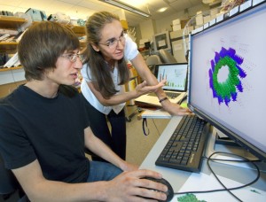
Gregory Alushin and Eva Nogales led a team of researchers that produced images of microtubule assembly and disassembly at the unprecedented resolution of 5 angstroms. (Photo by Roy Kaltschmidt)
During mitosis, the process by which a dividing cell duplicates its chromosomes and distributes them between two daughter cells, microtubules disassemble and reform into spindles across which the duplicate sets of chromosomes migrate. For chromosome migration to occur, the microtubules attached to them must disassemble, carrying the chromosomes in the process. 

The crucial ability of microtubules to transition from a rigid polymerized or “assembled” state to a flexible depolymerized or “disassembled” state – called “dynamic instability” – is driven by GTP hydrolysis in the microtubule lattice. Taxol prevents or dramatically slows down the unchecked cell division that is cancer by binding to a microtubule in such a manner as to block the effects of hydrolysis. However, until now the atomic details as to how microtubules transition from polymerized to depolymerized structures and the role that Taxol can play have been sketchy.

“Uncovering the atomic details of the conformational cycle accompanying polymerization, nucleotide hydrolysis, and depolymerization is essential for a complete description of microtubule dynamics,” Nogales says. “Such details should significantly aid in improving the potency and selectivity of existing anti-cancer drugs, as well as facilitate the development of novel agents.”

To find these details, Nogales, an expert in electron microscopy and image analysis and a leading authority on the structure and dynamics of microtubules, employed cryo-EM, in which protein samples are flash-frozen at liquid nitrogen temperatures to preserve their natural structure. Using an FEI 300 kV Titan cryo-EM from the laboratory of Robert Glaeser, she and her colleagues generated cryo-EM reconstructions of tubulin proteins whose structures were either stabilized by GMPCPP, a GTP analogue, or were unstable and bound to guanosine diphosphate (GDP), or were bound to GDP but stabilized by the presence of Taxol.
Alushin-Fig7_revised
The tubulin protein is a heterodimer consisting of alpha (α) and beta (β) monomer subunits. It features two guanine nucleotide binding sites, an “N-site” on the α-tubulin that is buried, and an “E-site” on the β-tubulin that is exposed when the tubulin is depolymerized. Previous microtubule reconstruction studies were unable to distinguish the highly similar α-tubulin and β-tubulin from each other.

“To be able to distinguish the α-tubulin from the β-tubulin, we had to resolve our images at better than 8 Å, which most prior cryo-EM studies were unable to do,” Nogales says. “For that, we marked the subunits with kinesin, a protein motor that distinguishes between α- and β-tubulin.”
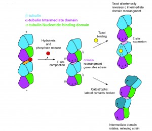
Nogales and her colleagues found that GTP hydrolysis and the release of the phosphate (GTP becomes GDP) leads to a compaction of the E-site and a rearrangement of the α-tubulin monomer that generates a strain on the microtubule that destabilizes its structure. Taxol binding leads to a reversal of this E-site compaction and α-tubulin rearrangement that restores structural stabilization.

“Remarkably, Taxol binding globally reverses the majority of the conformational changes we observe when comparing the GMPCPP and GDP states,” Nogales says. “We propose that GTP hydrolysis leads to conformational strain in the microtubule that would be released by bending during depolymerization. This model is consistent with the changes we observe upon taxol binding, which dramatically stabilizes the microtubule lattice. Our analysis supports a model in which microtubule-stabilizing agents like Taxol modulate conformational strain and longitudinal contacts in the microtubule lattice.”
This research was supported by NIH’s National Institute of General Medical Sciences, the Damon Runyon Cancer Research Foundation, and the Howard Hughes Medical Institute.

http://newscenter.lbl.gov/news-releases/2014/05/22/new-details-on-microtubules/

Wednesday, February 5, 2014

It’s the Water: Graphene Balloon Yields Unprecedented Images of Hydrated Protein Molecules

In this image generated by an electron microscope,
the white dots are the protein ferritin. The dark
circle in the middle is a bubble of liquid trapped within
 the graphene capsule enclosing the sample.
A graphene water balloon may soon open up new vistas for scientists seeking to understand health and disease at the most fundamental level.
Electron microscopes already provide amazingly clear images of samples just a few nanometers across. But if you want a good look at living tissue, look again.
“You can’t put liquid in an electron microscope,” says Tolou Shokuhfar, of Michigan Technological University. “So, if you have a hydrated sample—and all living things are hydrated—you have to freeze it, like a blueberry in an ice cube, and cut it into a million thin pieces, so the electrons can pass through. Only then can you image it to see what’s going on.”
After such treatment, the blueberry isn’t what it was, and neither is human tissue. Shokuhfar, an assistant professor of mechanical engineering-engineering mechanics, wondered if there might be a way to make electron microscopes more friendly to biological samples. That way, you might get a much better view of what’s really going on at the sub-cellular level.
So she joined colleagues at the University of Illinois-Chicago (UIC), and together they found a way. “You don’t need to freeze the blueberry, you don’t need to slice it up with a diamond knife,” she said. “You just put it in the electron microscope, and you can get down and see the atoms.”
The trick was to encapsulate the sample so that all the water stayed put while the electrons passed through freely. To do that, the team, including Robert F. Klie, an associate professor of physics and mechanical and industrial engineering at UIC, and UIC graduate student Canhui Wang, turned to graphene.
“Graphene is just a single layer of carbon atoms, and electrons can go through it easily, but water does not,” Klie said. “If you put a drop of water on graphene and top it with graphene, it forms this little balloon of water.” The graphene is strong enough to hold the water inside, even within the vacuum of an electron microscope.
The team tried their technique on a biochemical that plays a major role in human health: ferritin. “It’s a protein that stores and releases iron, which is critical for many body functions, and if ferritin isn’t working right, it may be contributing to lots of diseases, including Alzheimer’s and cancer,” Shokuhfar said.
The team made a microscopic sandwich, with ferritin immersed in water as the filling and graphene as the bread, and sealed the edges. Then, using a scanning transmission electron microscope, they captured a variety of images showing ferritin’s atomic structure.  In addition, they used a special type of spectroscopy to identify various atomic and electronic structures within the ferritin. Those images showed that the ferritin was releasing iron and pinpointed its specific form.
If the technique were used to compare ferritin taken from diseased tissue with healthy ferritin, it could provide new insights into illness at the molecular level. Those discoveries could lead to new treatments. “I believe this will allow us to identify disease signatures in ferritin and many other proteins,” Shokuhfar said.
An article on their work, “High-Resolution Electron Microscopy and Spectroscopy of Ferritin in Biocompatible Graphene Liquid Cells and Graphene Sandwiches,”  was published Feb. 4 online in Advanced Materials. Qiao Qiao, formerly a graduate student in Klie's UIC lab and now a postdoctoral fellow at Vanderbilt University, is also a coauthor on the study.
The work was funded by Michigan Technological University with additional support from a National Science Foundation grant to UIC, number DMR-0959470. The research was conducted at the University of Illinois-Chicago.
Source: http://www.mtu.edu/news/stories/2014/february/story102518.html

Thursday, January 16, 2014

Proteins Constantly Vibrate in the Body

(Image: healthy human T-cell; Credit NIAID/NIH)
The millions of proteins in humans and other living things vibrate in different patterns like the strings on a violin or the pipes of an organ, according to a new study in Nature Communications.
Scientists have long suspected that proteins vibrate in such a manner, but now they have the high tech means to prove that this really happens.
The research team, from the University at Buffalo and Hauptman-Woodward Medical Research Institute, found that the vibrations persist in molecules like the “ringing of a bell,” lead author and UB physics professor Andrea Markelz said in a press release.
We are not consciously aware of these non-stop vibrations. (Can you imagine what it would be like if we were?) But it’s fascinating to think that a veritable symphony of vibrations plays on in us and in other species.
The tiny motions enable proteins to change shape quickly so they can readily bind to other proteins, a process that is necessary for the body to perform critical biological functions like absorbing oxygen, repairing cells and replicating DNA, Markelz explained.
She added that the research opens the door to a whole new way of studying the basic cellular processes that enable life.
“People have been trying to measure these vibrations in proteins for many, many years, since the 1960s,” Markelz said.
She and her team managed to do it based on an interesting characteristic of proteins. They vibrate at the same frequency as the light they absorb. This is analogous to the way wine glasses tremble and shatter when a singer hits exactly the right note.
“Wine glasses vibrate because they are absorbing the energy of sound waves, and the shape of a glass determines what pitches of sound it can absorb,” she said. “Similarly, proteins with different structures will absorb and vibrate in response to light of different frequencies.”
In order to study vibrations in a protein known as “lysozyme,” the scientists then exposed a sample to light of different frequencies and polarizations, and measured the types of light the protein absorbed. This allowed them to identify which sections of the protein vibrated under normal biological conditions. The researchers were also able to see that the vibrations endured over time, challenging existing assumptions.
“If you tap on a bell, it rings for some time, and with a sound that is specific to the bell,” Markelz said. “This is how the proteins behave. Many scientists have previously thought a protein is more like a wet sponge than a bell: If you tap on a wet sponge, you don’t get any sustained sound.”
She concluded, “The cellular system is just amazing. You can think of a cell as a little machine that does lots of different things — it senses, it makes more of itself, it reads and replicates DNA, and for all of these things to occur, proteins have to vibrate and interact with one another.”

Source: http://news.discovery.com/human/health/life-hums-proteins-vibrate-in-body-140116.htm#mkcpgn=rssnws1

Wednesday, January 15, 2014

‘Make-or-break’ protein holds key to cancer spread

A potential cancer cell extruding from an epithelium. Courtesy of
Selwin Wu, Yap Lab, UQ's Institute for Molecular Bioscience
University of Queensland researchers have discovered a protein in cells that could block the escape route of potentially cancerous cells and stop them spreading to other parts of the body.

A team of biologists, physicists and mathematicians led by Professor Alpha Yap from UQ’s Institute for Molecular Bioscience made the discovery using microscopic imaging and statistical techniques.
“The finding could lead to new targeted treatments for cancer and other diseases,” Professor Yap said.
The researchers revealed and analysed molecular processes that cause potentially cancer-causing cells to escape from epithelial tissues, the layers of cells that cover and protect organs, including skin.
Professor Yap said the team had made important new insights into cancer biology, pinpointing the pathway these cells take to exit the epithelial tissue and investigating how the protein N-WASP can block their escape route.
“Abnormal or dying cells pose a risk to the health of the protective barrier that cells form around our organs,” he said.
“The normal cells that surround these dangerous cells use the complex process of cellular extrusion to push them out of the tissue,” Professor Yap said.
“However, when cancer cells are pushed out, it gives them the opportunity to grow or invade surrounding healthy tissue, which can cause the cancer to spread to other parts of the body and make it harder to control and treat.
“So while our normal cells think they’re doing us a favour by pushing out the bad cells, they’re actually helping the cancer cells to spread,” he said.
Professor Yap said his team had found a way to potentially block the escape routes by inhibiting the protein N-WASP, which regulates the internal skeleton of the cells.
“The pathway that makes or breaks these cells from escaping is regulated by N-WASP,” he said.
“We have found that if we can inhibit N-WASP from functioning, then we can stop these potentially cancerous cells from spreading.”
The study was conducted by researchers from UQ’s Institute for Molecular Bioscience in collaboration with Dr Zoltan Neufeld from UQ’s School of Mathematics and Physics.
The research has been published in the scientific journal Nature Cell Biology and was supported by the National Health and Medical Research Council of Australia, the Australian Research Council, the Kids Cancer Project of The Oncology Children’s Foundation, and The University of Queensland.
Confocal and optical microscopy was performed at the IMB’s ACRF Cancer Biology Imaging Facility, established with the generous support of the Australian Cancer Research Foundation.
The Institute for Molecular Bioscience (IMB) is a research institute of The University of Queensland that aims to improve quality of life by advancing medical genomics, drug discovery and biotechnology.
The UQ School of Mathematics and Physics has an international reputation for cutting-edge research and innovative teaching in mathematics, physics and statistics.


Tuesday, December 17, 2013

Alzheimer-substance may be the nanomaterial of tomorrow

It causes brain diseases like Alzheimer’s, Parkinson’s and Creutzfeldt-Jakob’s disease. It is also hard and rigid as steel. Now research at Chalmers University of Technology shows that the amyloid protein carries unique characteristics that may lead to the development of new composite materials for nano processors and data storage of tomorrow and even make objects invisible.

Piotr Hanczyc, PhD student at the department of Chemical and Biological Engineering, shows in an article in Nature Photonics, that the amyloid, a very dense aggregate of protein that causes brain diseases like Alzheimer's and Parkinson's, carries unique characteristics. Unlike well-functioning protein the amyloid reacts upon multi photon laser irradiation. This laser may in the future possibly be used for detection of amyloids inside a human brain. This discovery is in itself a breakthrough.

- But you can also create these aggregates in an artificial way in a laboratory and in combination with other materials create unique characteristics, Piotr Hanczyc says.

The amyloid aggregates are as hard and rigid as steel. The difference is that steel is much heavier and has defined material properties whereas amyloids can be tuned for desired purpose. By attaching a material’s molecules to the dense amyloid its characteristics change. This has been known for more than ten years and is already used by scientists.

- What hasn’t been known is that the amyloids react to multi-photon irradiation and this opens up new possibilities to also change the nature of the material attached to the amyloids, Piotr Hanczyc says.

The amyloids are shaped like discs densely piled upon each other.  When a material gets merged with these discs its molecules end up so densely and regularly that they can communicate and exchange information. This means totally new possibilities to change a material’s characteristics. 

Multi-photon tests on materials tied to amyloids are yet to be performed, but Piotr sees an opportunity for cooperation with Chalmers material science researchers interested for example in solar cell technology. 

And though it may still be science fiction, he also considers that one day scientists may use the material properties of amyloid fibrils in the research of invisible metamaterials.

- An object’s ability to reflect light could be altered so that what’s behind it gets reflected instead of the object itself, in principle changing the index of light refraction, kind of like when light hits the surface of water, Piotr Hanczyc says. 

Wednesday, November 13, 2013

Structure of bacterial nanowire protein hints at secrets of conduction

In this computer reconstruction done at EMSL, multiple 

Geobacter pilin structures are overlaid on Gonorrhea's 
fiber. Certain aromatic residues (either circled in light 
red or dangling out like threads) pop out of the fiber 
and provide insights into electrical conduction.
Tiny electrical wires protrude from some bacteria and contribute to rock and dirt formation. Researchers studying the protein that makes up one such wire have determined the protein's structure. The finding is important to such diverse fields as producing energy, recycling Earth's carbon and miniaturizing computers.
"This is the first atomic resolution structure of this protein from an electrically conductive bacterial species, and it sets the foundation for understanding how these nanowires work," said structural biologist Patrick Reardon of the Department of Energy's Pacific Northwest National Laboratory. Reardon is the 2012 William R. Wiley Distinguished Postdoctoral Fellow at EMSL, the DOE's Environmental Molecular Sciences Laboratory at PNNL.
With the help of related structures on disease-causing bacteria, the researchers show that the protein's shape and form suggest possible ways for the bacteria to shuttle electrons along the nanowire. The results were reported in October in the Journal of Biological Chemistry.
"How to get electrons from the inside of bacteria to the outside is important for many different things, such as bacterial fuel cells, how carbon cycles through the environment and how to make new nanomaterials for applications like biocomputers," said Reardon.
Aromatic Therapy
Many bacterial species wave fingerlike projections along their bodies. The bacteria use these fingers, called pili, to adhere to surfaces or weave into films or recognize objects in the environment. A group of related bacteria makes these bendy, stretchy structures out of a protein called pilin, and an even smaller group uses these structures like electrical wires.
Researchers and engineers would like to take advantage of this wiring. Bacteria produce electrons while respiring and use the wires to run electrons out of their little bacterial bodies. Normally the electrons build up or break down minerals in rock, but the system can also be used to clean up toxic heavy metals or to run a bacterial fuel cell.
To better understand how pilins contribute to conduction, Reardon and NMR lead scientist Karl Mueller explored pilin from an electrically conducting bacteria known as Geobacter sulfurreducens.
Previous research on Geobacter's pilin — PilA — provided a big hint. PilA required certain spots along its length known as aromatic residues to conduct electricity. Without those aromatic residues where they were, Geobacter had no zip in its pili.
But proteins are like a long string that folds up into a compact three-dimensional shape. Without knowing the shape of pilin, it wasn't clear where the aromatic residues landed in space or how they contributed to electron shuttling.
Hop or Flow?
To find out, the researchers used NMR — a technology similar to medical MRIs — at EMSL to picture the shape of PilA.
On its own, PilA looks like a long skinny spring, with a slight kink about halfway up. The aromatic residues, which are bulky anyway, bulge along its length. But the protein by itself isn't enough to reveal how conduction works. Many pilin proteins work together to form a fiber, and Reardon and Mueller only had one.
Nor did the researchers have the whole fiber to put into the NMR instrument. To get more clues, Reardon borrowed the computer image of an assembled fiber from an unrelated species, the bacteria that cause gonorrhea. Gonorrhea's fiber does not conduct electricity nor does its pilin have as many aromatic residues. But its pilin has a similar shape to PilA, so using a computer program, Reardon overlaid PilA on its Gonorrhea cousins.
At this point, the aromatic residues clearly stood out.
"We get clusters of aromatic residues, and they wrap along the wire candy cane style," said Reardon.
But that just raised another question. If the electrons traveling along Geobacter's pilin are using these aromatic residues, they could be hopping from aromatic island to aromatic island. Alternatively, the aromatic residues could be close enough to pass the electrons through like a baton in a running race. Reardon and Mueller agree the single structure is not enough to choose between the two options.
The next step, Mueller said, is to purify the whole fiber from Geobacter microbes and determine the complete structure. The task is technologically challenging however because the fiber has to be grown within the bacteria themselves. Visualizing the whole fiber, though, will show the scientists if the fiber resembles islands in a stream more, or the streambed itself.
This work was supported by the Department of Energy's Office of Science.

Monday, October 28, 2013

Berkeley Lab Researchers Get a Detailed Look at a DNA Repair Protein in Action


Errors in the human genetic code that arise from mismatched nucleotide base pairs in the DNA double helix can lead to cancer and other disorders. In microbes, such errors provide the basis for adaption to environmental stress. As one of the first responders to these genetic errors, a small protein called MutS – for “Mutator S” – controls the integrity of genomes across a wide range of organisms, from microbes to humans. Understanding the repair process holds importance for an equally impressive range of applications, including synthetic biology, microbial adaption and pathogenesis.


A new and detailed look at the role of MutS in DNA’s mismatch repair (MMR) system has been provided by a team of researchers with the U.S Department of Energy (DOE)’s Lawrence Berkeley National Laboratory (Berkeley Lab) and the Scripps Research Institute with their invention of a new technique for studying DNA. This breakthrough, which involves hybrid nanomaterials and small angle X-ray scattering (SAXS) technology, has been used to solve a major problem involving genome integrity and the biological detection of mismatched DNA.

Working at Berkeley Lab’s Advanced Light Source, the researchers used gold nanocrystal labels on DNA to create hybrid nanomaterials that are optimized for SAXS observation. The combination of gold-nanolabels and SAXS allowed the research team to follow DNA conformational changes brought on by MutS during the process of DNA mismatch error detection and response. They then showed that this hybrid nanolabel technique can also be used to examine short or long pieces of DNA in solutions that are comparable to cellular environments.

“Our technique of employing SAXS with gold nanolabels allows us to examine DNA processing by cooperative enzymes in which solution conditions, long distances, low concentrations, substoichiometric populations, and short time-scales are of importance,” says Greg Hura, a scientist with Berkeley Lab’s Physical Biosciences Division.

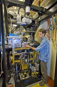
Greg Hura at the ALS SIBYLS beamline, which features two interchangeable end stations, one for macromolecular crystallography and one for small angle X-ray scattering. (Photo by Roy Kaltschmidt)
Greg Hura at the ALS SIBYLS beamline, which features endstations for macromolecular crystallography and small angle X-ray scattering. (Photo by Roy Kaltschmidt)
Hura is the lead author of a paper describing this research in the Proceedings of the National Academy of Sciences (PNAS). The article is titled “DNA conformations in mismatch repair probed in solution by X-ray scattering from gold nanocrystals.” The corresponding author is John Tainer, who holds joint appointments with Berkeley Lab’s Life Sciences Division and the Scripps Research Institute. (See below for complete list of co-authors)

“It is a common belief that DNA is a passive component in protein interactions that involve DNA metabolism, but many proteins actually make use of DNA structural features, such as rigidity and conformation for important biological processes,” Tainer says. “The view of DNA as a passive element is at least in part due to a paucity of robust tools for examining dynamic DNA conformational states during multistep reactions.”

For this study, Tainer, Hura and their colleagues were able to capitalize on the high quality X-ray beams at ALS beamline 12.3.1, also known as SIBYLS, which stands for Structurally Integrated Biology for Life Sciences. Maintained by Berkeley Lab’s Life Sciences Division under the direction of Tainer, the SIBYLS experimental station is optimized for SAXS imaging, which provides global information on the conformations adopted by a population of macromolecules in almost any solution condition.

“Because X-rays scatter predominantly from electrons, the use of  gold nanocrystals provides extremely high contrast relative to organic molecules critical for biology,” Tainer says. “This is important because we can follow specific biological molecules in complex reactions and along pathways to understand how their changes in shape and assembly control the biological outcomes.”

John Tainer, who holds joint appointments with Berkeley Lab and the Scripps Research Institute, directs the experimental stations at the ALS SIBYLS beamline.
The SAXS study at the SIBYLS beamline validated what has been dubbed the “beads-on-a-string” model of DNA repair, in which MutS proteins are the beads and DNA is the string. In solution, the MutS protein will bind to a mismatched DNA site by bending the DNA. ATP enzymes will come in to encircle and excise the error. The MutS then straightens out the bend and continues to proofread the DNA.

“This is the first time we used this technique to look at a protein-mediated process like DNA repair in solution with multiple partners,” Hura says. “We were able to determine some important details about MutS and the MMR system that should be valuable for drug design. We also now know what to look for in cancer-causing mutations of MutS. When we look at mutant versions of MutS we may be able to see that they do not bend the DNA or form the filament to the same extent as the normal version.”

In addition to Hura and Tainer, other co-authors of the PNAS paper were Chi-Lin Tsai, Shelley Claridge, Marc Mendilloc, Jessica Smith, Gareth Williams, Alexander Mastroianni, Paul Alivisatos, Christopher Putnam and Richard Kolodner.
This research was supported by the DOE Office of Science, the National Institutes of Health, and by the Berkeley Laboratory Directed Research and Development program.

Thursday, October 24, 2013

3D structure reveals protein’s Swiss-army knife strategy

The molecular machine that makes essential components of ribosomes – the cell’s protein factories – is like a Swiss-army knife, researchers at the European Molecular Biology Laboratory (EMBL) in Heidelberg, Germany, and the Centro de Investigaciones Biológicas in Madrid, Spain, have found. By determining the 3-dimensional structure of this machine, called RNA polymerase I, for the first time, the scientists found that it incorporates modules which prevent it from having to recruit outside help. The findings, published online today in Nature, can help explain why this protein works faster than its better-studied counterpart, RNA polymerase II. 
“Rather than recruiting certain components from outside, RNA polymerase I has them already built in, which explains why it is bigger, and less regulated, but at the same time more efficient,” says Christoph Müller from EMBL, who led the study. “Because everything is already assembled, there’s no time delay,” explains Maria Moreno-Morcillo, who carried out the work. 
There are three different RNA polymerases, each of which makes specific types of RNA molecule. For example, RNA polymerase II makes messenger RNA – the ‘middle-man’ that carries the information encoded in DNA to a ribosome where it can be used to make a protein. RNA polymerases I and III make parts of the machinery which reads that messenger RNA: I builds the RNA that will eventually form a ribosome, while III makes the transfer RNA that carries the protein building blocks to the ribosome for assembly. Scientists have known for over a decade what RNA polymerase II looks like and how it works, but obtaining detailed information on the structures of its counterparts has proven extremely difficult. Now that they have managed to do so for RNA polymerase I, Müller and colleagues have found explanations for some of the protein’s particularities.
Part of the difficulty in studying RNA polymerase I is that it is a larger molecule than RNA polymerase II. When they determined its 3-dimensional structure, the scientists found that some of the ‘extra’ modules in RNA polymerase I are remarkably similar to other, separate proteins that RNA polymerase II needs to do its job. It seems that RNA polymerase I has brought those helper modules permanently on board. In another part of the molecule, Müller and colleagues found that RNA polymerase I appears to have combined what in RNA polymerase II are two separate modules into a single, multi-tasking component. Together, these changes likely explain why RNA polymerase I can produce RNA molecules at a faster rate than RNA polymerase II.
The findings also imply that the cell has fewer ways of controlling RNA polymerase I’s activity, since it can’t influence it by changing the availability of helper proteins as it does in the case of RNA polymerase II. But here, too, RNA polymerase I’s Swiss-army knife strategy provides a solution. The structure showed that this molecular machine has a built-in regulatory mechanism: it can stop itself from attaching to DNA by bending a loop in its structure to block the space the DNA would usually dock onto.
The work was carried out in collaboration with Carlos Fernández-Tornero’s lab at the Centro de Investigaciones Biológicas in Madrid, Spain, as well as researchers at the University of Gӧttingen, Germany and the SOLEIL synchrotron in France, where some of the structural data was obtained. Structural data was also obtained at the Petra III ring at EMBL Hamburg, on the DESY campus in Germany.

Christoph Müller recently received an Advanced Grant from the European Research Council (ERC) to study RNA polymerase I and the proteins it interacts with.

Source Article

Fernández-Tornero, C., Moreno-Morcillo, M., Rashid, U.J., Taylor, N.M.I, Ruiz, F.M., Gruene, T., Legrand, P., Steuerwald, U. & Müller, C.W. Crystal structure of the 14-subunit RNA polymerase I. Published online in Nature on 23 October 2013. DOI: 10.1038/nature12636.

Article Abstract

Protein biosynthesis depends on the availability of ribosomes, which in turn relies on ribosomal RNA production. In eukaryotes, this process is carried out by RNA polymerase I (Pol I), a 14-subunit enzyme, whose activity is a major determinant of cell growth. Here, we present the crystal structure of Pol I fromSaccharomyces cerevisiae at 3.0 Å resolutionThe Pol I structure shows a compact core with a wide DNA-binding cleft and a tightly anchored stalk. An extended loop mimics the DNA backbone in the cleft and may be involved in regulating Pol I transcription. Subunit A12.2 extends from the A190 jaw to the active site and inserts a TFIIS-like zinc ribbon into the nucleotide triphosphate entry pore, providing insight into the role of A12.2 in RNA cleavage and Pol I insensitivity to ␣-amanitin. The A49/A34.5 heterodimer embraces subunit A135 through extended arms thereby contacting and potentially regulating subunit A12.2.

Thursday, October 10, 2013

Direct ‘writing’ of artificial cell membranes on graphene

Graphene emerges as a versatile new surface to assemble model cell membranes mimicking those in the human body, with potential for applications in sensors for understanding biological processes, disease detection and drug screening.

Writing in Nature Communications, researchers at The University of Manchester led by Dr Aravind Vijayaraghavan, and Dr Michael Hirtz at the Karlsruhe Institute of Technology (KIT), have demonstrated that membranes can be directly ‘written’ on to a graphene surface using a technique known as Lipid Dip-Pen Nanolithography (L-DPN).

The human body contains 100 trillion cells, each of which is enveloped in a cell membrane which is essentially a phospholipid bi-layer membrane. These cell membranes have a plethora of proteins, ion channels and other molecules embedded in them, each performing vital functions.

It is essential, therefore, to study and understand these systems, thereby enabling their application in areas such as bio-sensing, bio-catalysis and drug-delivery. Considering that it is difficult to accomplish this by studying live cells inside the human body, scientists have developed model cell membranes on surfaces outside the body, to study the systems and processes under more convenient and accessible conditions.

Dr Vijayaraghavan’s team at Manchester and their collaborators at KIT have shown that graphene is an exciting new surface on which to assemble these model membranes, and brings many advantages compared to existing surfaces.

Dr Vijayaraghavan  said: “Firstly, the lipids spread uniformly on graphene to form high-quality membranes. Graphene has unique electronic properties; it is a semi-metal with tuneable conductivity.

“When the lipids contain binding sites such as the enzyme called biotin, we show that it actively binds with a protein called streptavidin. Also, when we use charged lipids, there is charge transfer from the lipids into graphene which changes the doping level in graphene. All of these together can be exploited to produce new types of graphene/lipids based bio-sensors.”

Dr. Michael Hirtz (KIT) explains the L-DPN technique: “The technique utilizes a very sharp tip with an apex in the range of several nanometers as a means to write lipid membranes onto surfaces in a way similar to what a quill pen does with ink on paper. The small size of the tip and the precision machine controlling it allows of course for much smaller patterns, smaller than cells, and even right down to the nanoscale.”

“By employing arrays of these tips multiple different mixtures of lipids can be written in parallel, allowing for sub-cellular sized patterns with diverse chemical composition.”
 

Source: http://www.manchester.ac.uk/aboutus/news/display/?id=10831

Thursday, August 22, 2013

Researchers use an unnatural amino acid to design a novel metal-binding protein

Someday designer metal-binding proteins could catalyze chemical reactions unseen in nature for industrial and medical applications. Unfortunately, these proteins are difficult to create from scratch. Now, using computational design, researchers have generated a novel metal-binding protein by incorporating an unnatural amino acid with a taste for metals (J. Am. Chem. Soc. 2013, DOI: 10.1021/ja403503m).
None of the 20 amino acids typically found in proteins has a strong affinity for metals, says Jeremy H. Mills, a postdoctoral researcher in the laboratory of David Baker at the University of Washington, Seattle. So, metal-binding sites in natural proteins often contain several amino acids working together to grasp a metal. Mimicking that precise orientation of several amino acids poses a serious challenge in the design of metal-binding proteins, Mills says.
Baker, Mills, and their colleagues sidestepped the problem using an unnatural amino acid called (2,2’-bipyridin-5-yl)alanine, or Bpy-Ala, which has micromolar affinities for a variety of metals. At first, the team attempted to design an easy-to-express enzyme that mimicked the activity of catechol dioxygenase. This enzyme breaks down the environmental pollutant catechol, but is difficult to express in laboratories, Mills says.
They used computer programs to redesign catechol dioxygenase’s metal-binding site to include Bpy-Ala. Then, they searched a database of protein structures for ones that express well in Escherichia coli and could spatially accommodate the redesigned metal-binding site. With some small structural tweaks, the researchers identified five promising protein sequences.
After they expressed those designed proteins in E. coli, the researchers tested their ability to bind iron via a spectroscopic technique. Two of the proteins appeared to bind the metal, but when the researchers solved the structure of one using X-ray crystallography, they did not see the metal-binding site. Instead, the part of the protein containing the Bpy-Ala amino acid had twisted so that the unnatural amino acid jutted into solution. With the unnatural amino acid in the wrong position, the protein couldn’t catalyze the desired reaction.
Disappointed, the researchers set a humbler goal: design a protein that just binds metals. This time, they instructed their computer program to place Bpy-Ala within a more rigid part of the protein, such as within an α-helix. Such structures, according to Mills, “are less free to move around,” and more likely to lock the Bpy-Ala into place.
The strategy worked. They identified one easily expressed protein with about 250 amino acids that could bind iron, based on its spectroscopic signature. The crystal structure of the new protein matched their computer design almost exactly. Also the novel protein bound cobalt, zinc, and nickel, with affinities in the micromolar to picomolar range.
The match between the team’s designed structure and the X-ray crystal structure is impressive, says Huimin Zhao of theUniversity of Illinois, Urbana-Champaign, as well as their use of an unnatural amino acid to create the metal binding site. The next step, he says, is to design a metalloprotein “that catalyzes a reaction that a natural protein cannot.”