Showing posts with label human cell. Show all posts

Thursday, December 17, 2015

Scientists discover how cancer cells escape blood vessels


Study offers new targets for drugs that may prevent cancer from spreading.

Scientists at MIT and Massachusetts General Hospital have discovered how cancer cells latch onto blood vessels and invade tissues to form new tumors — a finding that could help them develop drugs that inhibit this process and prevent cancers from metastasizing.

Cancer cells circulating in the bloodstream can stick to blood vessel walls and construct tiny “bridges” through which they inject genetic material that transforms the endothelial cells lining the blood vessels, making them much more hospitable to additional cancer cells, according to the new study.

The researchers also found that they could greatly reduce metastasis in mice by inhibiting the formation of these nanobridges.

“Endothelial cells line every blood vessel and are the first cells in contact with any blood-borne element. They serve as the gateway into and out of tumors and have been the focus of intense research in vascular and cancer biology. These findings bring these two fields together to add greater insight into control of cancer and metastases,” says Elazer Edelman, the Thomas D. and Virginia W. Cabot Professor of Health Sciences and Technology, a member of MIT’s Institute for Medical Engineering and Science and of the Koch Institute for Integrative Cancer Research, and one of the leaders of the research team.

The lead author of the paper, which appears in the Dec. 16 issue of Nature Communications, is Yamicia Connor, a graduate student in the Harvard-MIT Division of Health Sciences and Technology (HST). The paper’s senior author is Shiladitya Sengupta, an assistant professor at HST and at Harvard Medical School.


Building bridges

Metastasis is a multistep process that allows cancer to spread from its original site and form new tumors elsewhere in the body. Certain cancers tend to metastasize to specific locations; for example, lung tumors tend to spread to the brain, and breast tumors to the liver and bone.

To metastasize, tumor cells must first become mobile so they can detach from the initial tumor.
Then they break into nearby blood vessels so they can flow through the body, where they become circulating tumor cells (CTCs). These CTCs must then find a spot where they can latch onto the blood vessel walls and penetrate into adjacent tissue to form a new tumor.

Blood vessels are lined with endothelial cells, which are typically resistant to intruders.

“Normal endothelial cells should not enable a cancer cell to invade, but if a cancer cell can connect with an endothelial cell, and inject signals that enable this endothelial cell to be controlled and completely transformed, then it facilitates metastasis,” Sengupta says.

The researchers first spotted tiny bridges between cancer cells and endothelial cells while using electron microscopy to study the interactions between those cell types. They speculated that the cancer cells might be sending some kind of signal to the endothelial cells.

“Once we saw that these structures allowed for a ubiquitous transfer of a lot of different materials, microRNAs were an obvious interesting molecule because they’re able to very broadly control the genome of a cell in ways that we don’t really understand,” Connor says. “That became our focus.”

MicroRNA, discovered in the early 1990s, helps a cell to fine-tune its gene expression. These strands of RNA, about 22 base pairs long, can interfere with messenger RNA, preventing it from being translated into proteins.

In this case, the researchers found, the injected microRNA makes the endothelial cells “sticky.”
That is, the cells begin to express proteins on their surfaces that attract other cells to adhere to them. This allows additional CTCs to bind to the same site and penetrate through the vessels into the adjacent tissue, forming a new tumor.

“It’s almost like the cancer cells are cooperating with each other to facilitate migration,” Sengupta says. “You just need maybe 1 percent of the endothelial cells to become sticky, and that’s good enough to facilitate metastasis.”

Non-metastatic cancer cells did not produce these invasive nanobridges when grown on endothelial cells.

Erkki Ruoslahti, a professor of cell, molecular, and developmental biology at the University of California at Santa Barbara, says that the discovery is an important advance in understanding tumor metastasis.

“I found it particularly interesting that the transfer of regulatory macromolecules from tumor cells to endothelial cells via intercellular nanotubes appears to be more effective (at least over relatively short distances) than exosome-mediated transfer, which has received a lot of attention lately,” says Ruoslahti, who was not part of the research team.

Shutting down metastasis

The nanobridges are made from the proteins actin and tubulin, which also form the cytoskeleton that gives cells their structure. The researchers found that they could inhibit the formation of these nanobridges, which are about 300 microns long, by giving low doses of drugs that interfere with actin.

When the researchers gave these drugs to mice with tumors that normally metastasize, the tumors did not spread.

Sengupta’s lab is now trying to figure out the mechanism of nanobridge formation in more detail, with an eye toward developing drugs that act more specifically to inhibit the process.

“If we can first understand how these structures are formed, then we can try to design targeted therapies to inhibit their formation, which could be a promising new area for developing drugs that specifically target metastasis,” Connor says.

Monday, November 2, 2015

First Complete Pictures of Cells' DNA-Copying Machinery


The first-ever images of the protein complex that unwinds, splits, and copies double-stranded DNA reveal something rather different from the standard textbook view. The electron microscope images, created by scientists at the U.S. Department of Energy's Brookhaven National Laboratory with partners from Stony Brook University and Rockefeller University, offer new insight into how this molecular machinery functions, including new possibilities about its role in DNA "quality control" and cell differentiation. The images and implications are described in a paper published online by the journal Nature Structural & Molecular Biology, November 2, 2015.

"This work is a continuation of our long-standing research using electron microscopy to understand the mechanism of DNA replication, an essential function for every living cell," said Huilin Li, a biologist with a joint appointment at Brookhaven Lab and Stony Brook University.

"These new images show the fully assembled and fully activated 'helicase' protein complex—which encircles and separates the two strands of the DNA double helix as it passes through a central pore in the structure—and how the helicase coordinates with the two 'polymerase' enzymes that duplicate each strand to copy the genome."

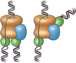
Three-dimensional structure of the active DNA helicase bound to the front-end DNA polymerase (Pol epsilon). The DNA polymerase epsilon (green) sits on top rather than the bottom of the helicase.


Studying this molecular machinery, known collectively as a "replisome," and the details of its DNA-copying process can help scientists understand what happens when DNA is miscopied—a major source of mutation that can lead to cancer—or learn more about how a single cell can eventually develop into the many cell types that make up a multicellular organism. But no one has produced a real structure of a replisome at any resolution for any organism until now.

"All the textbook drawings and descriptions of how a replisome should look and work are based on biochemical and genetic studies," Li said, likening the situation to the famous parable of the three blind men trying to describe an elephant, each looking at only one part. Those textbook drawings show the helicase moving along the DNA, separating the two strands of the double helix, with two polymerases located at the back where the DNA strand is split. In this configuration, the polymerases would add nucleotides (molecules containing the complementary A, T, G, and C bases of the genetic code) to the side-by-side split ends as they move out of the helicase to form two new complete double helix DNA strands.

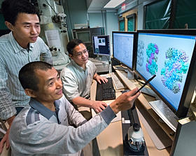
Collaborating scientists and study coauthors Zuanning Yuan, a graduate student at Stony Brook University (standing), Huilin Li of Stony Brook and Brookhaven Lab (seated, back), and Jingchuan Sun of Brookhaven Lab (seated, front) examining protein structures.

To test these assumptions, Li's group turned to the technique they had previously used to study individual components of the helicase, electron microscopy (EM). Jingchuan Sun, an EM expert in Li's lab, was essential to the success of the work. He studied samples of replisomes from baker's yeast cells—a model for all nucleus-containing cells—prepared and provided by Roxana Georgescu in Michael O'Donnell's research group at Rockefeller University. O'Donnell's group had previously published biochemical results related to this work.

"DNA replication is one of the most fundamental processes of life, so it is every biochemist's dream to see what a replisome looks like," Sun said. "Our lab has expertise and a decade of experience using electron microscopy to study DNA replication, which has prepared us well to tackle the highly mobile therefore very challenging replisome structure. Working together with the O'Donnell lab, which has done beautiful functional studies on the yeast replisome, our two groups brought perfectly complementary expertise to this project," he said.

The team's first-ever images of an intact replisome revealed that only one of the polymerases is located at the back of the helicase. The other is on the front side of the helicase, where the helicase first encounters the double-stranded helix. This means that while one of the two split DNA strands is acted on by the polymerase at the back end, the other has to thread itself back through or around the helicase to reach the front-side polymerase before having its new complementary strand assembled.

The scientists were so surprised by this finding that they asked another group at Rockefeller, led by Brian Chait, to perform additional structural studies using mass spectrometry. Yi Shi, a postdoctoral fellow in Chait's group performed this work, which confirmed the electron-microscopy-based conclusion about the unexpected architecture of the replisome.

The counterintuitive position of one polymerase at the front of the helicase suggests that it may have an unforeseen function. The authors suggest several possibilities including keeping the two "daughter" strands separate to help organize them during replication and cell division. It might also be possible that, as the single strand moves over other portions of the structure, some "surveillance" protein components check for lesions or mistakes in the nucleotide sequence before it gets copied—a sort of molecular quality control.

This architecture could also potentially play an important role in developmental biology by providing a pathway for treating the two daughter strands differently. Many modifications to DNA, including how it is packaged with other proteins, control which of the many genes in the sequence are eventually expressed in cells. An asymmetric replisome may result in asymmetric treatment of the two daughter strands during cell division—an essential step for making different tissues within a multicellular organism.

As the paper concludes, "Clearly, further studies will be required to understand the functional implications of the unexpected replisome architecture reported here."

This research was funded by the National Institutes of Health (GM103314, GM109824, GM74985, AG29979, and GM38839) and by the Howard Hughes Medical Institute.

Tuesday, September 15, 2015

Reconnect a severed spinal cord and repair paralysis using exosomes to hijack cell-to-cell communication


Regenerative medicine using stem cells is an increasingly promising approach to treat many types of injury. Transplanted stem cells can differentiate into just about any other kind of cell, including neurons to potentially reconnect a severed spinal cord and repair paralysis.

A variety of agents have been shown to induce transplanted stem cells to differentiate into neurons. Tufts University biomedical engineers recently published the first report of a promising new way to induce human mesenchymal stem cells (or hMSCs, which are derived from bone marrow) to differentiate into neuron-like cells: treating them with exosomes.

Exosomes are very small, hollow particles that are secreted from many types of cells. They contain functional proteins and genetic materials and serve as a vehicle for communication between cells. In the nervous system, exosomes guide the direction of nerve growth, control nerve connection and help regenerate peripheral nerves.

In a series of experiments reported in PLOS ONE in August, the Tufts researchers showed that exosomes from PC12 cells (neuron-like progenitor cells derived from rats) at various stages of their own differentiation could, in turn, cause hMSCs to become neuron-like cells. Exosomes had not previously been studied as a way to induce human stem cell differentiation.

The biomedical engineers also showed that the exosomes contain miRNAs—tiny pieces of RNA that regulate cell behavior and are known to play a role in neuronal differentiation. The researchers hypothesize that the exosomes caused the hMSCs to differentiate by delivering miRNA into the stem cells. The researchers plan future studies to determine the exact mechanism.

Friday, May 30, 2014

Hitchhiking nanotubes show how cells stir themselves

Rice, Göttingen, VU researchers track single-molecule proteins in living cells 

Chemical engineers from Rice University and biophysicists from Georg-August Universität Göttingen in Germany and the VU University Amsterdam in the Netherlands have successfully tracked single molecules inside living cells with carbon nanotubes.
Through this new method, the researchers found that cells stir their interiors using the same motor proteins that serve in muscle contraction.
The study, which sheds new light on biological transport mechanisms in cells, appears this week in Science.
The team attached carbon nanotubes to transport molecules known as kinesin motors to visualize and track them as they moved through the cytoplasm of living cells.
“I am amazed how versatile carbon nanotubes are,” said co-author Matteo Pasquali, a Rice professor of chemical and biomolecular engineering and of chemistry. “We use them for a wide range of applications, from engineering conducting fibers to imaging in cells.”
Carbon nanotubes are hollow cylinders of pure carbon with one-atom-thick walls. They naturally fluoresce with near-infrared wavelengths when exposed to visible light, a property discovered at Rice by Professor Rick Smalley a decade ago and then leveraged by Rice Professor Bruce Weisman to image carbon nanotubes. When attached to a molecule, the hitchhiking nanotubes serve as tiny beacons that can be precisely tracked over long periods of time to investigate small, random motions inside cells.
“Any probe that can hitch the length and breadth of the cell, rough it, slum it, struggle against terrible odds, win through and still know where its protein is, is clearly a probe to be reckoned with,” said lead author Nikta Fakhri, paraphrasing “The Hitchhiker’s Guide to the Galaxy.” Fakhri, who earned her Rice doctorate in Pasquali’s lab in 2011, is currently a Human Frontier Science Program Fellow at Göttingen.
“In fact, the exceptional stability of these probes made it possible to observe intracellular motions from times as short as milliseconds to as long as hours,” she said.
For long-distance transport, such as along the long axons of nerve cells, cells usually employ motor proteins tied to lipid vesicles, the cell’s “cargo containers.” This process involves considerable logistics: Cargo needs to be packed, attached to the motors and sent off in the right direction.
“This research has helped uncover an additional, much simpler mechanism for transport within the cell interior,” said principal investigator Christoph Schmidt, a professor of physics at Göttingen. “Cells vigorously stir themselves, much in the way a chemist would accelerate a reaction by shaking a test tube. This will help them to move objects around in the highly crowded cellular environment.”
The researchers showed the same type of motor protein used for muscle contraction is responsible for stirring. They reached this conclusion after exposing the cells to drugs that suppressed these specific motor proteins. The tests showed that the stirring was suppressed as well.
The mechanical cytoskeleton of cells consists of networks of protein filaments, like actin. Within the cell, the motor protein myosin forms bundles that actively contract the actin network for short periods. The researchers found random pinching of the elastic actin network by many myosin bundles resulted in the global internal stirring of the cell. Both actin and myosin play a similar role in muscle contraction. 
The highly accurate measurements of internal fluctuations in the cells were explained in a theoretical model developed by VU co-author Fred MacKintosh, who used the elastic properties of the cytoskeleton and the force-generation characteristics of the motors.
“The new discovery not only promotes our understanding of cell dynamics, but also points to interesting possibilities in designing ‘active’ technical materials,” said Fakhri, who will soon join the Massachusetts Institute of Technology faculty as an assistant professor of physics. “Imagine a microscopic biomedical device that mixes tiny samples of blood with reagents to detect disease or smart filters that separate squishy from rigid materials.”
Co-authors of the study include graduate student Alok Wessel, technical assistant Charlotte Willms and research scientist Dieter Klopfenstein, all of the University of Göttingen.
The German Research Foundation, the Dutch Foundation for Fundamental Research on Matter, the Netherlands Organization for Scientific Research, the Welch Foundation, the National Science Foundation and the Human Frontier Science Program supported the research.
-30-

View a short movie of nanotube-tagged proteins moving via stirring inside cells:


A thin carbon nanotube is attached to a molecular motor (yellow) that moves along microtubule filaments (green) that form the transport network of cells. This transport occurs in the highly crowded environment of the cytoplasm that includes a network of actin filaments (red). The fluorescent nanotube serves as a beacon for both the transport along the microtubule, as well as the buffeting of the microtubule by the highly agitated surrounding cytoplasm. (Credit: M. Leunissen, Dutch Data Design)


- See more at: http://news.rice.edu/2014/05/29/hitchhiking-nanotubes-show-how-cells-stir-themselves/#sthash.cQhqzdGm.dpuf

Monday, October 7, 2013


Gene activity and transcript patterns visualized for the first time in thousands of single human cells

Biologists of the University of Zurich have developed a method to visualize the activity of genes in single cells. The method is so efficient that, for the first time, a thousand genes can be studied in parallel in ten thousand single human cells. Applications lie in fields of basic research and medical diagnostics. The new method shows that the activity of genes, and the spatial organization of the resulting transcript molecules, strongly vary between single cells.

Whenever cells activate a gene, they produce gene specific transcript molecules, which make the function of the gene available to the cell. The measurement of gene activity is a routine activity in medical diagnostics, especially in cancer medicine. Today’s technologies determine the activity of genes by measuring the amount of transcript molecules. However, these technologies can neither measure the amount of transcript molecules of one thousand genes in ten thousand single cells, nor the spatial organization of transcript molecules within a single cell. The fully automated procedure, developed by biologists of the University of Zurich under the supervision of Prof. Lucas Pelkmans, allows, for the first time, a parallel measurement of the amount and spatial organization of single transcript molecules in ten thousands single cells. The results, which were recently published in the scientific journal Nature Methods, provide completely novel insights into the variability of gene activity of single cells.
Robots, a fluorescence microscope and a supercomputer

The method developed by Pelkmans’ PhD students Nico Battich and Thomas Stoeger is based upon the combination of robots, an automated fluorescence microscope and a supercomputer. “When genes become active, specific transcript molecules are produced. We can stain them with the help of a robot”, explains Stoeger. Subsequently, fluorescence microscope images of brightly glowing transcript molecules are generated. Those images were analyzed with the supercomputer Brutus, of the ETH Zurich. With this method, one thousand human genes can be studied in ten thousand single cells. According to Pelkmans, the advantages of this method are the high number of single cells and the possibility to study, for the first time, the spatial organization of the transcript molecules of many genes.
New insights into the spatial organization of transcript molecules

The analysis of the new data shows that individual cells distinguish themselves in the activity of their genes. While the scientists had been suspecting a high variability in the amount of transcript molecules, they were surprised to discover a strong variability in the spatial organization of transcript molecules within single cells and between multiple single cells. The transcript molecules adapted distinctive patterns.

“We realized that genes with a similar function also have a similar variability in the transcript patterns,” explains Battich. “This similarity exceeds the variability in the amount of transcript molecules, and allows us to predict the function of individual genes.” The scientists suspect that transcript patterns are a countermeasure against the variability in the amount of transcript molecules. Thus, such patterns would be responsible for the robustness of processes within a cell.

The importance of these new insights was summarized by Pelkmans: “Our method will be of importance to basic research and the understanding of cancer tumors because it allows us to map the activity of genes within single tumor cells.”
Source: http://www.mediadesk.uzh.ch/articles/2013/transkriptmuster_en.html

Wednesday, September 11, 2013

UAlberta researchers discover how immune system kills healthy cells

Basic science discovery could lead to improved treatments for cancer and viral infections.
Medical scientists at the University of Alberta have discovered how the immune system kills healthy cells while attacking infections. Their findings could one day lead to better treatments for cancer and viral infections.
Colin Anderson, a researcher with the Faculty of Medicine & Dentistry, recently published his team’s findings in the peer-reviewed Journal of Immunology. His team included colleagues from the United States and the Netherlands, and graduate students from the U of A.
Previous research has shown that when the immune system launches an aggressive attack on infected cells, healthy tissues and cells can be killed or damaged in the process. Anderson and his team discovered the mechanisms in the immune system that cause this “overkill” response.
“This opens the opportunity that one might be able to manipulate the immune-system response to block collateral damage without blocking the killing of infected cells,” Anderson explained.
“In the future, this might be important in the development of clinical treatments in cases where the immune system response needs to be harnessed. For example, in treating various viral infections, the collateral damage caused during the immune-system attack is a large part of the illness.
“In other cases, such as cancer or tumour treatments, one may want to increase the immune system’s ability to kill collateral cells, in hopes of killing tumour cells that would otherwise escape during treatment and spread elsewhere in the body. Our research suggests there are other mechanisms that could improve cancer therapy and make it more efficacious. This finding could also help us understand why certain cancer treatments are more successful than others.”
Anderson’s team discovered that “the weaponry the immune system uses to try to kill an infected or cancerous cell is not exactly the same as the weaponry that causes collateral damage to innocent bystander cells that aren’t infected.” For years, it was assumed the weaponry to kill infected cells versus healthy cells was exactly the same.
The research group is continuing work in this area to see whether they can alter the level of collateral damage to healthy cells without altering the attack on infected cells.
Anderson is a researcher in the Department of Surgery and the Department of Medical Microbiology and Immunology. He is also a member of the Alberta Diabetes Institute and the Alberta Transplant Institute.
The research was funded by the Canadian Institutes of Health Research.
Source: http://news.ualberta.ca/newsarticles/2013/september/ualberta-researchers-discover-how-immune-system-kills-healthy-cells?utm_source=feedburner&utm_medium=feed&utm_campaign=Feed%3A+UofAExpressNewsArticles+%28University+of+Alberta+News%29#sthash.GFhgdxPH.dpuf