Showing posts with label cells. Show all posts

Wednesday, May 14, 2014

BYU researchers create tiny nano-device in newest gene therapy advance

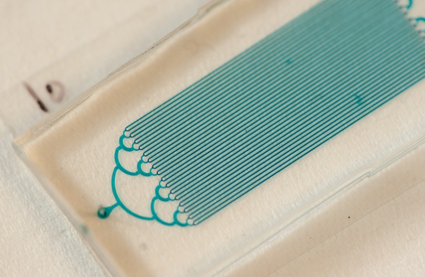
This SEM (scanning electron microscope) image shows the nanoinjector next to a latex bead the same size as an egg cell. You can see the size of the nanoinjector and its lance compared to a cell.
Credit: Brian Jensen/BYU

Nanoinjector is used to transfer genes and DNA to new cells

The ability to transfer a gene or DNA sequence from one animal into the genome of another plays a critical role in the medical research of diseases such as cancer, Alzheimer’s and diabetes.

But the traditional method of transferring genetic material into a new cell, microinjection, has a serious downside. This method uses a hollow needle to pump a DNA-filled liquid into an egg cell nucleus, but that extra fluid causes the cell to swell and die 40 percent of the time.

Now a multidisciplinary team of Brigham Young University scientists has developed a way to significantly reduce cell death when introducing DNA into egg cells. The researchers have created a microscopic lance that delivers DNA to the cells through electrical forces.

“Because DNA is naturally negatively charged, it is attracted to the outside of the lance using positive voltage,” said Brian Jensen, BYU professor of mechanical engineering. “Once we insert the lance into a cell, we simply reverse the polarity of the electrical force and the lance releases the DNA.”

Because the lance is 10 times smaller and no extra fluid is used, the cells undergo significantly less stress compared to microinjection, and thus, have a higher survival rate. The researchers describe their “metamorphic nanoinjection” process in an article published today by Review of Scientific Instruments.

Currently the BYU researchers, which include microbiology professor Sandra Burnett and mechanical engineering professor Larry Howell, are using the technique to inject DNA into mouse zygotes (single-cell embryos consisting of a fertilized egg).

“The microinjection technology hasn’t really changed over the last 40-50 years since it was invented,” Burnett said. “Not having to force liquid into the nuclei by shifting to a lance is a huge advantage. It not only increases the survival rate, but it also causes less damage for future development.”

In research published in Transgenic Research, the team found that 77.6% of nanoinjected mouse zygotes proceeded to the two-cell stage of development as compared to 54.7% for microinjected zygotes.

A major reason for creating transgenic animals is to research genetic or infectious diseases. By modifying the genes of a mouse to carry a human disease, researchers can generate data with insights into future treatments and therapies for those illnesses.

One of the BYU team’s most significant findings is that it’s possible to use the electrical forces to get DNA into the nucleus of a cell without aiming the lance into the pronucleus (the cellular structure containing the cell’s DNA). This may mean that injections can be performed in animals with cloudy or opaque embryos.

“Such animals, including many interesting larger ones like pigs, would be attractive for a variety of transgenic technologies,” Jensen said. “We believe nanoinjection may open new fields of discovery in these animals.”

Jensen said more efficient injections should also reduce the cost to create transgenic animals. Jensen’s research is funded in part by the $400,000 awarded to him in 2011 through a National Science Foundation CAREER Award.

Quentin Aten, a former PhD student at BYU now with Nexus Spine, LLC, served as the lead author on the research published in Review of Scientific Instruments.

View the study here:http://scitation.aip.org/content/aip/journal/rsi/85/5/10.1063/1.4872077


http://news.byu.edu/archive14-may-nanoinjector.aspx

Wednesday, April 9, 2014

UC San Diego Researchers Develop Bacterial ‘FM Radio’

Programming living cells offers the prospect of harnessing sophisticated biological machinery for transformative applications in energy, agriculture, water remediation and medicine.  Inspired by engineering, researchers in the emerging field of synthetic biology have designed a tool box of small genetic components that act as intracellular switches, logic gates, counters and oscillators.
But scientists have found it difficult to wire the components together to form larger circuits that can function as “genetic programs.”  One of the biggest obstacles? Dealing with a small number of available wires.
A team of biologists and engineers at UC San Diego has taken a large step toward overcoming this obstacle. Their advance, detailed in a paper which appears in this week’s advance online publication of the journal Nature, describes their development of a rapid and tunable post-translational coupling for genetic circuits. This advance builds on their development of “biopixel” sensor arrays reported in Nature by the same group of scientists two years ago. 
The problem the researchers solved arises from the noisy cellular environment that tends to lead to highly variable circuit performance. The components of a cell are intermixed, crowded and constantly bumping into each other. This makes it difficult to reuse parts in different parts of a program, limiting the total number of available parts and wires. These difficulties hindered the creation of genetic programs that can read the cellular environment and react with the execution of a sequence of instructions.
The team’s breakthrough involves a form of “frequency multiplexing” inspired by FM radio.
“This circuit lets us encode multiple independent environmental inputs into a single time series,” said Arthur Prindle, a bioengineering graduate student at UC San Diego and the first author of the study. “Multiple pieces of information are transferred using the same part. It works by using distinct frequencies to transmit different signals on a common channel.”
The key that enabled this breakthrough is the use of frequency, rather than amplitude, to convey information. “Combining two biological signals using amplitude is difficult because measurements of amplitude involve fluorescence and are usually relative. It’s not easy to separate out the contribution of each signal,” said Prindle. “When we use frequency, these relative measurements are made with respect to time, and can be readily extracted by measuring the time between peaks using any one of several analytical methods.”
While their application may be inspired by electronics, the UC San Diego scientists caution in their paper against what they see as increasing “metaphorization” of engineering biology.
“We explicitly make the point that since biology is often too intertwined to engineer in the way we are accustomed in electronics, we must deal directly with bidirectional coupling and quantitatively understand its effects using computational models,” explained Prindle. “It’s important to find the right dose of inspiration from engineering concepts while making sure you aren’t being too reliant on your engineering metaphors.”
Microfluidic device containing an array of "biopixels" (square traps) in which bacteria grow. Credit: Arthur Prindle, UC San Diego
Enabling this breakthrough is the development of an intracellular wiring mechanism that enables rapid transmission of protein signals between the individual modules. The new wiring mechanism was inspired by a previous study in the lab on the bacterial stress response. It reduces the time lags that develop as a consequence of using proteins to activate or repress genes.
“The new coupling method is capable of reducing the signaling time delay between individual genetic circuits by more than an order of magnitude,” said Jeff Hasty, a professor of biology and bioengineering at UC San Diego who headed the team of researchers and co-directs the university’s BioCircuits Institute. “The state of the art has been about 20 to 40 minutes, but we can now do it in less than one minute.”
“This study is an outstanding example of the power of interdisciplinary systems biology approaches, which treat our cells like integrated pathways and networks instead of a collection of individual components,” said Sarah Dunsmore, a program manager at the National Institute of General Medical Sciences, which finances a National Center for Systems Biology at UC San Diego that supported the research. “By combining the complexity of naturally occurring biological processes with engineering principles, Dr. Hasty and colleagues have produced a model that will provide the basis for creating genetic circuits that can be used to study human health and disease.”
“What’s really exciting about this coupling method is the particular way we did it,” said Prindle. “Rather than trying to build from scratch, we made use of the enzyme machinery that the cell uses for rapid and precise signaling during times of stress. This is an appealing strategy because it lets us take advantage of the advanced machinery that nature has already evolved.”
Hasty credited Prindle for coming up with the idea for the study and carrying it through. “Beyond his modeling and bench skills, I’ve been extremely impressed by Arthur’s ingenuity and drive,” said Hasty. “This project arose from his creativity at the outset and he had the raw energy and excitement to carry it to the end.”
The scientists received funding for their development from the National Science Foundation (MCB-1121748) and by the San Diego Center for Systems Biology (NIH Grant P50 GM085764).

http://ucsdnews.ucsd.edu/pressrelease/uc_san_diego_researchers_develop_bacterial_fm_radio#When:17:00:00Z

Monday, February 10, 2014

Nanomotors Are Controlled, for the First Time, Inside Living Cells

For the first time, a team of chemists and engineers at Penn State University have placed tiny synthetic motors inside live human cells, propelled them with ultrasonic waves and steered them magnetically. It's not exactly "Fantastic Voyage," but it's close. The nanomotors, which are rocket-shaped metal particles, move around inside the cells, spinning and battering against the cell membrane.
"As these nanomotors move around and bump into structures inside the cells, the live cells show internal mechanical responses that no one has seen before," said Tom Mallouk, Evan Pugh Professor of Materials Chemistry and Physics at Penn State. "This research is a vivid demonstration that it may be possible to use synthetic nanomotors to study cell biology in new ways. We might be able to use nanomotors to treat cancer and other diseases by mechanically manipulating cells from the inside. Nanomotors could perform intracellular surgery and deliver drugs noninvasively to living tissues."
The researchers' findings will be published in Angewandte Chemie International Edition on 10 February 2014. In addition to Mallouk, co-authors include Penn State researchers Wei WangSixing LiSuzanne Ahmed, and Tony Jun Huang, as well as Lamar Mairof Weinberg Medical Physics in Maryland U.S.A.
The interaction between gold nanorods and HeLa cells in acoustic fields showing strong attachment. Videos play at original speed, and were taken under 500Xoverall magnification in dark field. Duration: 17 seconds. Credit: Mallouk Lab, Penn State University.
Up until now, Mallouk said, nanomotors have been studied only "in vitro" in a laboratory apparatus, not in living human cells. Chemically powered nanomotors first were developed ten years ago at Penn State by a team that included chemistAyusman Sen and physicist Vincent Crespi, in addition to Mallouk. "Our first-generation motors required toxic fuels and they would not move in biological fluid, so we couldn't study them in human cells," Mallouk said. "That limitation was a serious problem." When Mallouk and French physicistMauricio Hoyos discovered that nanomotors could be powered by ultrasonic waves, the door was open to studying the motors in living systems.
The assembly of a rotating HeLa cell/gold rod aggregate at an acoustic nodal line in the xy plane. The video was taken under 500X overall magnification except for 00:23 - 00:32 and 01:16 - 01:42, where a 200X overall magnification was used. Duration: 2:49. Credit: Mallouk Lab, Penn State University.
 
For their experiments, the team uses HeLa cells, an immortal line of human cervical cancer cells that typically is used in research studies. These cells ingest the nanomotors, which then move around within the cell tissue, powered by ultrasonic waves. At low ultrasonic power, Mallouk explained, the nanomotors have little effect on the cells. But when the power is increased, the nanomotors spring into action, moving around and bumping into organelles -- structures within a cell that perform specific functions. The nanomotors can act as egg beaters to essentially homogenize the cell's contents, or they can act as battering rams to actually puncture the cell membrane.
A demonstration of very active gold nanorods internalized inside HeLa cells in an acoustic field. This video was taken under 1000X magnification in the bright field, with most of the incoming light blocked at the aperture. Credit: Mallouk Lab, Penn State University.
While ultrasound pulses control whether the nanomotors spin around or whether they move forward, the researchers can control the motors even further by steering them, using magnetic forces. Mallouk and his colleagues also found that the nanomotors can move autonomously -- independently of one another -- an ability that is important for future applications. "Autonomous motion might help nanomotors selectively destroy the cells that engulf them," Mallouk said. "If you want these motors to seek out and destroy cancer cells, for example, it's better to have them move independently. You don't want a whole mass of them going in one direction."
The ability of nanomotors to affect living cells holds promise for medicine, Mallouk said. "One dream application of ours is Fantastic Voyage-style medicine, where nanomotors would cruise around inside the body, communicating with each other and performing various kinds of diagnoses and therapy. There are lots of applications for controlling particles on this small scale, and understanding how it works is what's driving us."
The research was funded by the National Science Foundation (MRSECgrant DMR-0820404), the National Institutes of Health, the Huck Innovative and Transformative Seed Fund (HITS) and Penn State University.

CONTACTS
Tom Mallouk: tem5@psu.edu, (+1) 814-863-9637
Barbara Kennedy (PIO): science@psu.edu, (+1) 814-863-4682

Source: http://science.psu.edu/news-and-events/2014-news/Mallouk2-2014

Friday, February 7, 2014

Growing brains in the lab

Human embryonic stem cells spontaneously organize
into neuroepithelial tissue containing multiple zones
after growing for 70 days in culture.
© 2014 Yoshiki Sasai, RIKEN Center for
Developmental Biology
Human embryonic stem cells can be induced to spontaneously form developing brain tissue

During development, the nervous system forms as a flat sheet called the neuroepithelium on the outer layer of the embryo. This sheet eventually folds in on itself to form a neural tube that gives rise to the brain and spinal cord—a process that involves the proliferation and migration of immature nerve cells to form the brain at one end and the spinal cord at the other. Yoshiki Sasai, Taisuke Kadoshima and colleagues from the RIKEN Center for Developmental Biology have now shown that human embryonic stem (ES) cells can spontaneously organize into the cerebral cortical tissue that forms at the front, or ‘brain’ end, of the developing neural tube
Sasai and his colleagues previously developed a novel cell culture technique that involves growing ES cells in suspension, and have shown that these cells can self-organize into complex three-dimensional structures. They have already used this method to grow pieces of cerebral cortex and embryonic eyes from mouse ES cells. And more recently, they have shown that human ES cells can also organize into embryonic eyes containing retinal tissue and light-sensitive cells.
In their most recent work, Sasai’s team treated human ES cells grown using their cell culture system with signaling molecules that induce the formation of nervous tissue from the outer embryonic layer. They found that the cells spontaneously organize into neuroepithelial tissue that then folds up to give a multilayered cortex (Fig. 1).
During human embryonic development, the neural tube thickens at both ends. In particular, the front end thickens dramatically as waves of cells migrate outward to form the layered cerebral cortex and other parts of the brain. An important finding of the team’s is that the front end of the neural tube appears to thicken due to the growth of radial glial fibers, which span the thickness of the tube and guide migrating cells, rather than due to the accumulation of immature cells within the tube, as previously thought. 
The findings also highlight critical differences between the development of the neural tube in mice and humans. While in humans, the inner surface of the neural tube and the intermediate neuroepithelial zone underneath it contain distinct populations of neural progenitors resembling radial glia, the progenitor population in the latter is not present in the developing mouse cortex.
“Efficient generation of cortical tissues could provide a valuable resource of functional neurons and tissues for medical applications,” says Kadoshima. “By combining this method with disease-specific human induced pluripotent stem cells, it will also be possible to reproduce complex human disorders.”
Source: http://www.rikenresearch.riken.jp/eng/research/7637

Tuesday, February 4, 2014

Scientists turn primitive artificial cell into complex biological materials


It is a big dream in science: To start from scratch with simple artificial microscopic building blocks and end up with something much more complex: living systems, novel computers or every-day materials. For decades scientists have pursued the dream of creating artificial building blocks that can self-assemble in large numbers and reassemble to take on new tasks or to remedy defects. Now researchers from University of Southern Denmark have taken a step forward to make this dream come true.

“The potential of such new man-made systems is almost limitless, and many expect these novel materials to become the foundation of future technologies”, says Dr. Maik Hadorn from Department of Chemistry and Applied Biosciences at ETH Zürich, who conducted the research as a postdoctoral research fellow at University of Southern Denmark (SDU).

Over the last three years he and the colleagues Eva Boenzli,Kristian T. Sørensen and Martin M. Hanczyc from the Center for Fundamental Living Technology (FLinT) at SDU have worked on the challenges of making primitive building blocks assemble and turn into something functional.

“We used short DNA strands as smart glue to link preliminary stages of artificial cells (called artificial vesicles) to engineer novel tissue-like structures”, says Dr. Maik Hadorn.

As part of the EU-sponsored project MATCHIT (MATrix for CHemical Information Technology) Dr. Maik Hadorn and coworkers have earlier showed that short DNA strands can guide the self-assembly process of artificial vesicles; that two types of artificial vesicles can be linked in a way predefined by the person conducting the experiment, and that assembled structures can be reassembled, when triggered externally.

In their most recent scientific article, published in Langmuir in December 2013, the researchers from SDU, in collaboration with colleagues from Italy and Japan, not only increased the complexity of the self-assembled structures that are now composed of several types of artificial vesicles – they also loaded one vesicle type with a basic cellular machinery derived from bacterial cells. This enabled these vesicles to translate an encapsulated genetic blueprint into a functional protein.

Put together the researchers have managed to engineer controlled assemblies that are visible to the naked eye and that resemble natural tissues in their architecture as well as in their functionalities.

Methods of constructing simple artificial structures have been known for decades, but only the use of DNA strands that act as a smart glue has allowed the researchers to overcome shortcomings of precedent methods and to engineer higher-order structures of predefined and programmable architecture.

“As the artificial vesicles resemble natural cells both in size and composition, they are an ideal starting point for a multitude of applications. One application can be a temporal support for wound healing: A wound may be covered with assemblies of vesicles that are tailored in a patient specific manner. They will not only protect the natural cells beneath the wound but also initiate and guide the differentiation of these cells so that they divide and differentiate. Finally, these regenerated natural cells can take over and fulfill their protective function”, explains Maik Hadorn.

The new systems are also of value in studying cells:

“Natural organisms are complex. Simple model systems like our tissue-like structures may help to reveal secrets for example of cell communication and cell differentiation”.

Besides these two potential applications in personalized medicine and natural sciences, one can also think of using assembled vesicles as small bioreactors.

“It’s somehow like cooking”, Dr. Hadorn explains:

“If you’re preparing a meal, most of the time you’re not using just one pot. To prepare your meat, potatoes, and also the vegetables in just one pot is almost impossible. By using different pots you’re making sure that the conditions for the preparation of each component are optimal and that the components only meet if they are ready. Transferred to current one-pot bioreactors in science we often face similar problems. However, by using microscopic pots (i.e. vesicles) that are loaded with a defined set of substances and that are in close proximity to one another, one can think of microscopic bioreactors in which gates open to release substances from one vesicle into a neighboring vesicles. This ensures that the reaction conditions are optimal for the synthesis of products too complex for today’s one-pot bioreactors.”


Source: http://sdu.dk/en/Om_SDU/Fakulteterne/Naturvidenskab/Nyheder/2014_02_04_guv_cells

Wednesday, December 18, 2013

Cells from the eye are inkjet printed for the first time

Close-up of retinal cells in a jet
IOP Publising, Biofabrication
A group of researchers from the UK have used inkjet printing technology to successfully print cells taken from the eye for the very first time.

The breakthrough, which has been detailed in a paper published today, 18 December, in IOP Publishing’s journal Biofabrication, could lead to the production of artificial tissue grafts made from the variety of cells found in the human retina and may aid in the search to cure blindness.
At the moment the results are preliminary and provide proof-of-principle that an inkjet printer can be used to print two types of cells from the retina of adult rats―ganglion cells and glial cells. This is the first time the technology has been used successfully to print mature central nervous system cells and the results showed that printed cells remained healthy and retained their ability to survive and grow in culture.
Co-authors of the study Professor Keith Martin and Dr Barbara Lorber, from the John van Geest Centre for Brain Repair, University of Cambridge, said: “The loss of nerve cells in the retina is a feature of many blinding eye diseases. The retina is an exquisitely organised structure where the precise arrangement of cells in relation to one another is critical for effective visual function”.
“Our study has shown, for the first time, that cells derived from the mature central nervous system, the eye, can be printed using a piezoelectric inkjet printer. Although our results are preliminary and much more work is still required, the aim is to develop this technology for use in retinal repair in the future.”
Printed glia cells
IOP Publising,
Biofabrication

The ability to arrange cells into highly defined patterns and structures has recently elevated the use of 3D printing in the biomedical sciences to create cell-based structures for use in regenerative medicine.
In their study, the researchers used a piezoelectric inkjet printer device that ejected the cells through a sub-millimetre diameter nozzle when a specific electrical pulse was applied. They also used high speed video technology to record the printing process with high resolution and optimised their procedures accordingly.
“In order for a fluid to print well from an inkjet print head, its properties, such as viscosity and surface tension, need to conform to a fairly narrow range of values. Adding cells to the fluid complicates its properties significantly,” commented Dr Wen-Kai Hsiao, another member of the team based at the Inkjet Research Centre in Cambridge.
Once printed, a number of tests were performed on each type of cell to see how many of the cells survived the process and how it affected their ability to survive and grow.
The cells derived from the retina of the rats were retinal ganglion cells, which transmit information from the eye to certain parts of the brain, and glial cells, which provide support and protection for neurons.
“We plan to extend this study to print other cells of the retina and to investigate if light-sensitive photoreceptors can be successfully printed using inkjet technology. In addition, we would like to further develop our printing process to be suitable for commercial, multi-nozzle print heads,” Professor Martin concluded.
The research was undertaken by Dr. Barbara Lorber, also at the John van Geest Centre for Brain Repair, in collaboration with Dr. Wen-Kai Hsiao and Prof. Ian Hutchings from the Inkjet Research Centre, University of Cambridge. The work was funded by Fight for Sight, the van Geest Foundation and the EPSRC.
From Wednesday 18 December, the paper can be downloaded fromhttp://iopscience.iop.org/1758-5090/6/1/015001/article

Tuesday, December 17, 2013

"Nanobiopsy" allows scientists to operate on living cells

Scientists have developed a device that can take a "biopsy" of a living cell, sampling minute volumes of its contents without killing it.
Much research on molecular biology is carried out on populations of cells, giving an average result that ignores the fact that every cell is different. Techniques for studying single cells usually destroy them, making it impossible to look at changes over time.
The new tool, called a nanobiopsy, uses a robotic glass nanopipette to pierce the cell membrane and extract a volume of around 50 femtolitres – 0.00000000000005 litres, around one per cent of the cell’s contents.
It will allow scientists to take samples repeatedly, to study the progression of disease at a molecular level in an individual cell. It can also be used to deliver material into cells, opening up ways to reprogram diseased cells.
“This is like doing surgery on individual cells,” said Dr Paolo Actis, from the Department of Medicine at Imperial College London, who developed the technology with colleagues at the University of California, Santa Cruz.
“This technology will be extremely useful for research in many areas. You could use it to dynamically study how cancer cells are different from healthy cells, or look at how brain cells are affected by Alzheimer’s disease. The possibilities are immense.”
To get inside the cell, the nanopipette is plunged downwards about one micrometre to pierce the cell membrane. Applying a voltage across the tip makes fluid flow into the pipette. When the pipette is removed from the cell, the membrane remains intact and the cell retains its shape.
The device is based on a scanning ion conductance microscope, which uses a robotic nanopipette, about 100 nanometres in diameter, to scan the surface of cells. The nanopipette is filled with an electrolyte solution and the ion current is measured inside the tip. When the pipette gets close to a cell membrane, the ion current decreases. This measurement is used to guide the tip across the surface of a sample at a constant distance, producing a picture of the surface.
In an initial study published in the journal ACS Nano, the researchers used the nanobiopsy technique to extract and sequence messenger RNA, molecules carrying genetic code transcribed from DNA in the cell’s nucleus. This allowed them to see which genes were being expressed in the cell.
They were also able to extract whole mitochondria – the power units of the cell. Mitochondria contain their own DNA, and the researchers discovered that the genomes of different mitochondria in the same cell are different.
They are now working on adapting the technology to incorporate sensors on the pipette tip that can instantly measure different molecules.
The research was funded by the US National Cancer Institute and National Institutes of Health.

Monday, November 25, 2013

Scientists capture 'redox moments' in living cells


Better understanding of hardy bacteria enhances tool for biofuel creation

Scientists have charted a significant signaling network in a tiny organism that's big in the world of biofuels research. The findings about how a remarkably fast-growing organism conducts its metabolic business bolster scientists' ability to create biofuels using the hardy microbe Synechococcus, which turns sunlight into useful energy.

The team at the Department of Energy's Pacific Northwest National Laboratory glimpsed key chemical events, known as redox reactions, inside living cells of the organism. The publication in ACS Chemical Biology marks the first time that redox activity, a very fast regulatory network involved in all major aspects of a cell's operation, has been observed in specific proteins within living cells.

The findings hone scientists' control over a common tool in the biofuels toolbox. At a more basic level, the work gives researchers the newfound ability to witness a basic biological process that occurs every moment in everything from bacteria to people.

"Redox activity tells us where the action is going on within a cell," said chemist Aaron Wright, the leader of the PNNL team whose project was funded by DOE's Office of Science. "We've been able to get a look at the redox system while it's still operating in a living cell, without destroying the cell first. This allows us to tell who the players are when the cells are engaged in the activity of our choice, like making components for biofuels."

Redox activity is one of the most powerful tools an organism has to sense and adapt to a changing environment; it's particularly active in plants that must respond constantly to changing conditions, such as light and dark.

The PNNL study was aimed at ferreting out proteins that are potential redox players in the cyanobacterium Synechococcus. Cyanobacteria absorb light energy from the sun and use it to convert carbon dioxide into food and other molecules, while also giving off oxygen. Redox reactions play a role in directing where the harvested energy goes.

Scientists believe the organism and its plant-like cousins, including algae, were responsible for producing the first oxygen on Earth, more than 2.5 billion years ago. It's a sure bet that you have inhaled oxygen molecules produced by Synechococcus, which today contributes a significant proportion of the oxygen available on Earth.

The organism is attractive to scientists for a number of reasons. It's adept at converting carbon dioxide into other molecules, such as fatty acids, that are of interest to energy researchers. Synechococcus is easy for scientists to change and manipulate as they explore new ideas. And it grows quickly, doubling in approximately two hours. A patch just two feet wide by seven feet long — roughly the area of a typical dining room table — could blossom into an area the size of a football field in just one day.

Biofuels makers and other scientists are trying to exploit this ability to churn out quantities of materials that might serve as biofuel. Synechococcus is also remarkably hardy, capable of tolerating the stress caused by intense sunlight, which kills many other cyanobacteria. Redox reactions that take place throughout the organism are at the core of this ability, and understanding them gives scientists a treasured global view of how the cell lives and responds to change.

Some researchers are working to get the bacteria itself to create biofuel, growing an organism with more fatty acids that could be converted to diesel fuel. Others, like Wright, are working to understand the organism more completely, to direct the organism to create fuels using light and carbon dioxide.

Wright's team found the signals by keeping the bacteria hungry, then suddenly flooding it with food — a massive, immediate change in environment. Within 30 seconds, the team detected redox activity, which changes the way proteins operate by adding or subtracting electrons.

His team uncovered an extensive network of redox activity, identifying 176 proteins that are sensitive to signaling in this manner. Before this study, just 75 of those proteins were known to be part of a redox signaling network. The scientists found that the system is involved in all the major processes of a cell — which genes are turned on and off, for example, as well as how the cell maintains its molecular machinery and converts energy into fuel.

Central to the work are the chemical probes Wright developed that are able to cross the cell membrane and get into the cytoplasm of the cell. The probes flag redox events by binding to certain forms of the amino acid cysteine, which is a known player in many of these interactions. Then the probes and the interactions they flag are subjected to scrutiny at EMSL, the DOE's Environmental Molecular Sciences Laboratory on the PNNL campus, where instruments detect redox activity through various means, such as through fluorescent imaging and mass spectrometry. The analysis tells scientists about when and where within the cell redox activity occurred.

"Knowing the proteins that are sensitive to redox signaling lets us know where to look as we test out new methods for working with this organism," said Wright. "We can tinker with a specific protein, for instance, and then watch the effects immediately.

"This is the type of information we really must have if we want organisms like this to produce substances that make a difference, like biofuels, chemicals or potential medicines," he added.


Source: http://www.pnnl.gov/news/release.aspx?id=1024

Wednesday, September 11, 2013

UAlberta researchers discover how immune system kills healthy cells

Basic science discovery could lead to improved treatments for cancer and viral infections.
Medical scientists at the University of Alberta have discovered how the immune system kills healthy cells while attacking infections. Their findings could one day lead to better treatments for cancer and viral infections.
Colin Anderson, a researcher with the Faculty of Medicine & Dentistry, recently published his team’s findings in the peer-reviewed Journal of Immunology. His team included colleagues from the United States and the Netherlands, and graduate students from the U of A.
Previous research has shown that when the immune system launches an aggressive attack on infected cells, healthy tissues and cells can be killed or damaged in the process. Anderson and his team discovered the mechanisms in the immune system that cause this “overkill” response.
“This opens the opportunity that one might be able to manipulate the immune-system response to block collateral damage without blocking the killing of infected cells,” Anderson explained.
“In the future, this might be important in the development of clinical treatments in cases where the immune system response needs to be harnessed. For example, in treating various viral infections, the collateral damage caused during the immune-system attack is a large part of the illness.
“In other cases, such as cancer or tumour treatments, one may want to increase the immune system’s ability to kill collateral cells, in hopes of killing tumour cells that would otherwise escape during treatment and spread elsewhere in the body. Our research suggests there are other mechanisms that could improve cancer therapy and make it more efficacious. This finding could also help us understand why certain cancer treatments are more successful than others.”
Anderson’s team discovered that “the weaponry the immune system uses to try to kill an infected or cancerous cell is not exactly the same as the weaponry that causes collateral damage to innocent bystander cells that aren’t infected.” For years, it was assumed the weaponry to kill infected cells versus healthy cells was exactly the same.
The research group is continuing work in this area to see whether they can alter the level of collateral damage to healthy cells without altering the attack on infected cells.
Anderson is a researcher in the Department of Surgery and the Department of Medical Microbiology and Immunology. He is also a member of the Alberta Diabetes Institute and the Alberta Transplant Institute.
The research was funded by the Canadian Institutes of Health Research.
Source: http://news.ualberta.ca/newsarticles/2013/september/ualberta-researchers-discover-how-immune-system-kills-healthy-cells?utm_source=feedburner&utm_medium=feed&utm_campaign=Feed%3A+UofAExpressNewsArticles+%28University+of+Alberta+News%29#sthash.GFhgdxPH.dpuf

Wednesday, August 21, 2013

An Organized Approach to 3D Tissue Engineering

IBN’s novel technique brings researchers closer to viable organ implants

Researchers at the Institute of Bioengineering and Nanotechnology (IBN) have developed a simple method of organizing cells and their microenvironments in hydrogel fibers. Their unique technology provides a feasible template for assembling complex structures, such as liver and fat tissues, as described in their recent publication in Nature Communications1.
According to IBN Executive Director Professor Jackie Y. Ying, “Our tissue engineering approach gives researchers great control and flexibility over the arrangement of individual cell types, making it possible to engineer prevascularized tissue constructs easily. This innovation brings us a step closer toward developing viable tissue or organ replacements.”
IBN Team Leader and Principal Research Scientist, Dr Andrew Wan, elaborated, “Critical to the success of an implant is its ability to rapidly integrate with the patient’s circulatory system. This is essential for the survival of cells within the implant, as it would ensure timely access to oxygen and essential nutrients, as well as the removal of metabolic waste products. Integration would also facilitate signaling between the cells and blood vessels, which is important for tissue development.”
Tissues designed with pre-formed vascular networks are known to promote rapid vascular integration with the host. Generally, prevascularization has been achieved by seeding or encapsulating endothelial cells, which line the interior surfaces of blood vessels, with other cell types. In many of these approaches, the eventual distribution of vessels within a thick structure is reliant on in vitro cellular infiltration and self-organization of the cell mixture. These are slow processes, often leading to a non-uniform network of vessels within the tissue. As vascular self-assembly requires a large concentration of endothelial cells, this method also severely restricts the number of other cells that may be co-cultured.
Alternatively, scientists have attempted to direct the distribution of newly formed vessels via three-dimensional (3D) co-patterning of endothelial cells with other cell types in a hydrogel. This approach allows large concentrations of endothelial cells to be positioned in specific regions within the tissue, leaving the rest of the construct available for other cell types. The hydrogel also acts as a reservoir of nutrients for the encapsulated cells. However, co-patterning multiple cell types within a hydrogel is not easy. Conventional techniques, such as micromolding and organ printing, are limited by slow cell assembly, large volumes of cell suspension, complicated multi-step processes and expensive instruments. These factors also make it difficult to scale up the production of implantable 3D cell-patterned constructs. To date, these approaches have been unsuccessful in achieving vascularization and mass transport through thick engineered tissues.
To overcome these limitations, IBN researchers have used interfacial polyelectrolyte complexation (IPC) fiber assembly, a unique cell patterning technology patented by IBN, to produce cell-laden hydrogel fibers under aqueous conditions at room temperature. Unlike other methods, IBN’s novel technique allows researchers to incorporate different cell types separately into different fibers, and these cell-laden fibers may then be assembled into more complex constructs with hierarchical tissue structures. In addition, IBN researchers are able to tailor the microenvironment for each cell type for optimal functionality by incorporating the appropriate factors, e.g. proteins, into the fibers. Using IPC fiber assembly, the researchers have engineered an endothelial vessel network, as well as cell-patterned fat and liver tissue constructs, which have successfully integrated with the host circulatory system in a mouse model and produced vascularized tissues.
The IBN researchers are now working on applying and further developing their technology toward engineering functional tissues and clinical applications.