Showing posts with label atomic. Show all posts

Monday, February 10, 2014

Massive neutrinos solve a cosmological conundrum

Scientists have solved a major problem with the current standard model of cosmology identified by combining results from the Planck spacecraft and measurements of gravitational lensing in order to deduce the mass of ghostly sub-atomic particles called neutrinos.

The team, from the universities of Manchester and Nottingham, used observations of the Big Bang and the curvature of space-time to accurately measure the mass of these elementary particles for the first time.
The recent Planck spacecraft observations of the Cosmic Microwave Background (CMB) – the fading glow of the Big Bang – highlighted a discrepancy between these cosmological results and the predictions from other types of observations.
The CMB is the oldest light in the Universe, and its study has allowed scientists to accurately measure cosmological parameters, such as the amount of matter in the Universe and its age. But an inconsistency arises when large-scale structures of the Universe, such as the distribution of galaxies, are observed.
Professor Richard Battye, from the University of Manchester’s School of Physics and Astronomy, said: “We observe fewer galaxy clusters than we would expect from the Planck results and there is a weaker signal from gravitational lensing of galaxies than the CMB would suggest.
“A possible way of resolving this discrepancy is for neutrinos to have mass. The effect of these massive neutrinos would be to suppress the growth of dense structures that lead to the formation of clusters of galaxies.”
Neutrinos interact very weakly with matter and so are extremely hard to study. They were originally thought to be massless but particle physics experiments have shown that neutrinos do indeed have mass and that there are several types, known as flavours by particle physicists. The sum of the masses of these different types has previously been suggested to lie above 0.06 eV (much less than a billionth of the mass of a proton).
In this paper, Professor Battye and co-author Dr Adam Moss, from the University of Nottingham, have combined the data from Planck with gravitational lensing observations in which images of galaxies are warped by the curvature of space-time. They conclude that the current discrepancies can be resolved if massive neutrinos are included in the standard cosmological model. They estimate that the sum of masses of neutrinos is 0.320 +/- 0.081 eV (assuming active neutrinos with three flavours).
Dr Moss said: “If this result is borne out by further analysis, it not only adds significantly to our understanding of the sub-atomic world studied by particle physicists, but it would also be an important extension to the standard model of cosmology which has been developed over the last decade.”
The paper is published in Physical Review Letters on 7 February and has been selected as an Editor’s choice.
Ends

Notes for editors

Jodrell Bank's role in Planck
This paper makes use of CMB data from the European Space Agency’s Planck spacecraft -http://www.esa.int/Our_Activities/Space_Science/Planck - and from the South Pole Telescope -http://pole.uchicago.edu/.
Jodrell Bank Centre for Astrophysics (JBCA - http://www.jodrellbank.manchester.ac.uk/) is directly involved with the two lowest frequencies of the Low Frequency Instrument on board Planck, the 30 and 44 GHz radiometers. These have four and six detectors respectively, operating at 20Kelvin (-253.15°C or -423.67°F). The resolution on the sky is 33 and 27 arc minutes, and the sensitivity 1.6 and 2.4 micro K (over 12 months). The cryogenic low noise amplifiers which are the heart of the radiometers were developed at Jodrell Bank, with help from the National Radio Astronomy Observatory in Virginia, USA.
Dr B Maffei and Dr G Pisano are involved in the other focal instrument, the HFI. First at Cardiff University and now at The University of Manchester, they have played a major role in the design, development and calibration of the Focal Plane Unit, in particular the cold optics, in collaboration with the Institut d'Astrophysique Spatiale, France, Maynooth University, Ireland and JPL/Caltech, USA.
The work to understand the Galactic emission seen by Planck is being co-led from Jodrell Bank by Emeritus Professor Rod Davies and Dr Clive Dickinson. A number of projects are led by Jodrell Bank scientists, including Professor Richard Davis and Dr Clive Dickinson. Each of the 14 projects focuses on one aspect of the Galaxy as seen by Planck, including the electrons that gyrate in the Galactic magnetic field, the ionized gas that pervades the interstellar medium and the dust grains that emit across the entire frequency range that Planck is sensitive to. Jodrell Bank is also leading the calibration and identifying systematics in the LFI data.
Jodrell Bank and Gravitational Lensing
This work uses gravitational lensing data from the Canada-France-Hawaii Telescope Lensing Survey (CFHTLenS - http://www.cfhtlens.org/).
The first gravitational lens to be discovered, the Double Quasar, was found by Dr Denis Walsh of The University of Manchester during a radio survey of the northern sky using telescopes at Jodrell Bank. Follow-up work by Walsh and collaborators using an optical telescope at Kitt Peak in the USA, led to its identification as the first gravitational lens (see Walsh et al 1979, Nature 279, 381). The quasar is at a redshift of 1.41 (a co-moving distance of about 13.7 billion light years) and is lensed by a bright galaxy (and associated cluster of galaxies) at a redshift of 0.355 (a co-moving distance of about 4.54 billion light years) forming a second image of the quasar core and inner jet.
One of the major current e-MERLIN  - http://www.e-merlin.ac.uk/ - legacy programmes (led by Neal Jackson of The University of Manchester and Stephen Serjeant of The Open University) will take advantage of its unique combination of sharpness of view and sensitivity to faint emission to map the multiple images in gravitational lenses and thereby study the evolution of the distribution of mass in distant galaxies.
For further information contact:
Aeron Haworth
Media Relations
Faculty of Engineering and Physical Sciences
The University of Manchester
Tel: 0161 275 8387
Mob: 07717 881563
Email: aeron.haworth@manchester.ac.uk

Source: http://www.manchester.ac.uk/aboutus/news/display/?id=11555

Wednesday, October 23, 2013

Atomic bonds in fullerene

A team of international researchers from the IBM in Zurich has published the first detailed single-molecule images of atomic bonds in fullerene.

According to the report published in Science, the new study has examined fullerene, which has linked rings of carbon atoms at its core, using an atomic force microscopy or AFM. 

Their images show the length of the atomic bonds. The bright and dark spots on the images correspond to the higher and lower density of electrons in the particle.
Atomic level imaging has come a long way in the past decade, and after scientists first managed to image molecular structure and even electron clouds, now a group of researchers at IBM Research Center Zurich have visually depicted how chemical bonds differentiate in individual molecules using a technique called non-contact atomic force microscopy (AFM).

In the image below one can clearly see detailed chemical bonds between individual atoms of a nanographene molecule or C60. In 3-D the molecule resembles a buckyball thanks to its football shape.

Atomic Bond

If you look closely you can see that some C-C chemical bonds are more highlighted than others. This is because in reality and practice, the  bonds between individual atoms differ slightly and subtly in length and strength, and for the first time we’ll now able to distinguish the different types of bonds from one another, visually.  The bright and dark spots correspond to higher and lower densities of electrons.

“In the case of pentacene, we saw the bonds but we couldn’t really differentiate them or see different properties of different bonds,” said lead author of the study Dr.  Leo Gross.
“Now we can really prove that… we can see different physical properties of different bonds, and that’s really exciting.”

Atomic Bond

The nanographene molecule imaged through the ATF versus the schematic of the molecule. (c) IBM Research Zurich
The nanographene molecule imaged through AFM versus the schematic of the molecule. (c) IBM Research Zurich

To create the images, the IBM researchers used an atomic force microscope with a tip that ended with a single carbon monoxide molecule. The CO molecule traces the image by oscillating between the tip and the sample. By measuring its wiggle and inter-molecular force  the AFM can slowly build up a very detailed image. The technique made it possible to distinguish individual bonds that differ by only three picometers, which is one-hundredth of an atom’s diameter.

“We found two different contrast mechanisms to distinguish bonds. The first one is based on small differences in the force measured above the bonds. We expected this kind of contrast but it was a challenge to resolve,” said IBM scientist Leo Gross. “The second contrast mechanism really came as a surprise: Bonds appeared with different lengths in AFM measurements. With the help of ab initio calculations we found that the tilting of the carbon monoxide molecule at the tip apex is the cause of this contrast.”

The findings were reported in the journal Science.

Read more at http://www.zmescience.com/research/individual-chemical-bonds-imaged-ibm-zurich-0123332/#isJ6kDRzO1BPtIoc.99

Thursday, October 10, 2013

Evidence for a new nuclear ‘magic number’

Researchers have come one step closer to understanding unstable atomic nuclei.  A team of researchers from RIKEN, the University of Tokyo and other institutions in Japan and Italy has provided evidence for a new nuclear magic number in the unstable, radioactive calcium isotope 54Ca. In a study published today in the journal Nature, they show that 54Ca is the first known nucleus with 34 neutrons (N) where N = 34 is a magic number.

The protons and neutrons inside the atomic nucleus exhibit shell structures in a manner similar to electrons in an atom. For naturally stable nuclei, these nuclear shells fill completely when the number of protons or the number of neutrons is equal to the ‘magic’ numbers 2, 8, 20, 28, 50, 82 or 126.
However, it has recently been shown that the traditional magic numbers, which were once thought to be robust and common for all nuclei, can in fact change in unstable, radioactive nuclei that have a large imbalance of protons and neutrons.
In the current study led by David Steppenbeck of the Center for Nuclear Study, the University of Tokyo, the team of researchers focused on 54Ca, which has 20 protons and 34 neutrons in its nucleus. They were able to study this nucleus thanks to the Radioactive Isotope Beam Factory (RIBF) at RIKEN, which produces the highest intensity radioactive beams available in the world.
In their experiment, a radioactive beam composed of scandium-55 and titanium-56 nuclei travelling at around 60% of the speed of light, was selected and purified by the BigRIPS fragment separator, part of the RIBF. The radioactive beam was focused on a reaction target made of beryllium. Inside this target, projectile fragmentation of the 55Sc and56Ti nuclei occurred, creating numerous new radioactive nuclei, some in excited states. The researchers measured the energy of the γ rays emitted from excited states of the radioactive nuclei using an array of 186 detectors surrounding the reaction target.
The results of the experiment indicate that 54Ca’s first excited state lies at a relatively high energy, which is characteristic of a large nuclear shell gap, thus indicating that N = 34 in 54Ca is a new magic number, as predicted theoretically by the University of Tokyo group in 2001. By conducting a more detailed comparison to nuclear theory the researchers were able to show that the N = 34 magic number is equally as significant as some other nuclear shell gaps.
“Our new measurement provides key data for the understanding of neutron-rich nuclei and will help pin down the treatment of nuclear forces in systems far from stability,” explains David Steppenbeck.
“Enriching our knowledge of the structures of highly unstable nuclei and the nucleon-nucleon forces that drive nuclear shell evolution and the appearance or disappearance of the nuclear magic numbers in radioactive nuclei plays an important role in understanding astrophysical processes such as nucleosynthesis in stars,” he adds.

Thursday, September 12, 2013

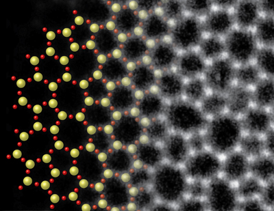
world's thinnest sheet of glass: one molecule thick

At just a molecule thick, it’s a new record: The world’s thinnest sheet of glass, a serendipitous discovery by scientists at Cornell and Germany’s University of Ulm, is recorded for posterity in the Guinness Book of World Records.

The “pane” of glass, so impossibly thin that its individual silicon and oxygen atoms are clearly visible via electron microscopy, was identified in the lab of David A. Muller, professor of applied and engineering physics and director of the Kavli Institute at Cornell for Nanoscale Science.

The work that describes direct imaging of this thin glass was published in January 2012 in Nano Letters, and the Guinness records officials took note. They published the achievement in early September for inclusion in the 2014 book, and the breakthrough is featured in the publication’s 21st Century Science spread.

Just two atoms in thickness, making it literally two-dimensional, the glass was an accidental discovery, Muller said. The scientists had been making graphene, a two-dimensional sheet of carbon atoms in a chicken wire crystal formation, on copper foils in a quartz furnace. They noticed some “muck” on the graphene, and upon further inspection, found it to be composed of the elements of everyday glass – silicon and oxygen.

Provided
An illustration shows a structural model, left, of 2-D glass, with electron microscopy data, right, of 2-D glass.

They concluded that an air leak had caused the copper to react with the quartz, also made of silicon and oxygen. This produced the glass layer on the would-be pure graphene.

Besides its sheer novelty, Muller continued, the work answers an 80-year-old question about the fundamental structure of glass. Scientists, with no way to directly see it, had struggled to understand it: It behaves like a solid but was thought to look more like a liquid.

Now, the Cornell scientists have produced a picture of individual atoms of glass, and they found it strikingly resembles a diagram drawn in 1932 by W.H. Zachariasen – a longstanding theoretical representation of the arrangement of atoms in glass.

“This is the work that, when I look back at my career, I will be most proud of,” Muller said. “It’s the first time that anyone has been able to see the arrangement of atoms in a glass.”

What’s more, two-dimensional glass could someday find a use in transistors, by providing a defect-free, ultra-thin material that could improve, for example, the performance of processors in computers and smartphones.

The paper, “Direct Imaging of a Two-Dimensional Silica Glass on Graphene,” was published in Nano Letters on Jan. 23, 2012, with first authors Pinshane Huang, a Cornell graduate student, and Simon Kurash, a University of Ulm graduate student. It includes collaborators from the University of Ulm, Germany; the Max Planck institute for Solid State Research in Germany; University of Vienna; University of Helsinki; and Aalto University in Finland.

The work at Cornell was funded by the National Science Foundation through the Cornell Center for Materials Research.


Thursday, August 22, 2013

Two become one with the 3D NanoChemiscope


Unique surface analysis instrument

The 3D NanoChemiscope is a miracle of state-of-the-art analysis technology. As a further development of well-known microscopic and mass spectroscopic methods, it maps the physical and chemical surfaces of materials down to the atomic level. This instrument, which is unique in the world, not only delivers high-definition images; it also knows what it is "seeing".

The result of a combined three-dimensional ToF-SIMS-/SFM surface analysis of a PCBM/CyI-polymer blend used by Empa's Functional Polymers Laboratory to produce organic solar cells.

What do a penguin and the surface of a solar cell have in common? Not a lot concedes Empa physicist Laetitia Bernard. Yet she must have smiled when, while processing an image of a polymer blend required to produce a new type of organic solar cell, at a certain point she could make out more and more clearly the outline of a penguin. A small detail in the complex world of high-performance microscopy. The 3D NanoChemiscope, which was developed at Empa, not only maps samples with nanometre precision, but for the first time can also provide precise information about which chemical elements are arranged where in a sample. This enables both mechanical properties, such as hardness, elasticity or friction, and chemical properties of surfaces to be determined simultaneously in three dimensions. In the case of the "penguin” image, this means that the 3D NanoChemiscope not only captures the outline of the "penguin", but also detects which polymers are located at its "beak", at its "eye" and "around" it. Using this analysis technique, the solar cell researchers are able to efficiently control the mechanisms of their materials and adapt the composition or concentration of their polymer blend accordingly. This enables new structures and therefore leads to better performances of the solar cell to be created.


Some of the many individual images from which the 3D NanoChemiscope generated the 3D view. 
The SFM scans the topography of the surface (The image on the left shows a section 12µm x 12µm in size. The differences in height visible in the image measure 100-200nm). 

With the TOF-SIMS, it is possible to identify where the different materials or polymers in the polymer blend are located on the surface (The images in the middle and on the right show C-+C2- and CN-+I- ions).

Scanning force microscope and high-end mass spectrometer

This analysis is made possible by the 3D NanoChemiscope, which combines two previously independent techniques. The scanning force microscope (SFM) scans the surface with an ultra-fine tip, while the time-of-flight secondary ion mass spectrometer (ToF-SIMS) determines the material composition of the first surface mono-layer by "shooting" metallic ions at it.

Up to now, in order to study both the chemical and physical properties of surfaces, it was necessary to analyse the sample in two different instruments. However, when transporting the sample from one instrument to the other, there was always a danger of contamination or oxidation. In addition, it was practically impossible to find the exact location scanned by the SFM again. What, therefore, could be more appropriate than to "combine" the two instruments? In a four-year project sponsored by the EU, project leader Laetitia Bernard, together with Empa researchers and partners from academia and industry, has carried out meticulous work to develop a new instrument in which an SFM and a ToF-SIMS are placed in an ultra-high vacuum chamber as near to each other as possible.


Mechanical engineer Sasa Vranjkovic and Laetitia Bernard, leader of the 3D NanoChemiscope project, discussing the construction drawing of a component.

The microscope experts have also equipped the 3D NanoChemiscope with a novel transport system developed in-house, which uses piezomotors to move the sample gently back and forth on tracks coated with a diamond-like carbon layer (DLC). The sample holder can move along five axes, allowing the location under investigation to be analysed from any angle.

Following its construction, the prototype – a monster made of gleaming aluminium 1 metre long, 70 centimetres wide and 1.7 metres tall – has been in operation at project partner ION-TOF GmbH in Münster, Germany, where it is being used by industrial clients and research partners. The construction of more instruments is planned, customers having expressed a keen interest and being prepared to pay sums over one million Swiss francs.


Wednesday, August 21, 2013

New Results from Daya Bay: Tracking the Disappearance of Ghostlike Neutrinos

Daya Bay neutrino experiment releases high-precision measurement of subatomic shape shifting and new result on differences among neutrino masses


The international Daya Bay Collaboration has announced new results about the transformations of neutrinos - elusive, ghostlike particles that carry invaluable clues about the makeup of the early universe.  The latest findings include the collaboration's first data on how neutrino oscillation – in which neutrinos mix and change into other "flavors," or types, as they travel – varies with neutrino energy, allowing the measurement of a key difference in neutrino masses known as "mass splitting."  
"Understanding the subtle details of neutrino oscillations and other properties of these shape-shifting particles may help resolve some of the deepest mysteries of our universe," said Jim Siegrist, Associate Director of Science for High Energy Physics at the U.S. Department of Energy (DOE), the primary funder of U.S. participation in Daya Bay. 
U.S. scientists have played essential roles in planning and running of the Daya Bay experiment, which is aimed at filling in the details of neutrino oscillations and mass hierarchy that will give scientists new ways to test for violations of fundamental symmetries. For example, if scientists detect differences in the way neutrinos and antineutrinos oscillate that are beyond expectations, it would be a sign of charge-parity (CP) violation, one of the necessary conditions that resulted in the predominance of matter over antimatter in the early universe.  The new results from the Daya Bay experiment about mass-splitting represent an important step towards understanding how neutrinos relate to the structure of our universe today.  
"Mass splitting represents the frequency of neutrino oscillation," says Kam-Biu Luk of the U.S. Department of Energy's Lawrence Berkeley National Laboratory (Berkeley Lab), the Daya Bay Collaboration's Co-spokesperson, who identified the ideal site for the experiment. "Mixing angles, another measure of oscillation, represent the amplitude. Both are crucial for understanding the nature of neutrinos." Luk is a senior scientist in Berkeley Lab's Physics Division and a professor of physics at the University of California (UC)  Berkeley.  
The Daya Bay Collaboration, which includes more than 200 scientists from six regions and countries, is led in the U.S. by DOE's  Berkeley Lab and Brookhaven National Laboratory (BNL). The Daya Bay Experiment is located close to the Daya Bay and Ling Ao nuclear power plants in China, 55 kilometers northeast of Hong Kong.  The latest results from the Daya Bay Collaboration will be announced at the XVth International Workshop on Neutrino Factories, Super Beams and Beta Beams in Beijing, China. 
"These new precision measurements are a great indication that our efforts will pay off with a deeper understanding of the structure of matter and the evolution of the universe – including why we have a universe made of matter at all," says Steve Kettell, a Senior Scientist at BNL and U.S. Daya Bay Chief Scientist. 
U.S. contributions to the Daya Bay experiment include coordinating detector engineering; perfecting the recipe for the liquid used to track neutrinos in the Daya Bay detectors; overseeing the photo-detector systems used to observe neutrino interactions and muons; building the liquid-holding acrylic vessels and the detector-filling and automated calibration systems; constructing the muon veto system; developing essential software and data analysis techniques; and managing the overall project. 

Measuring neutrino mass and flavors 

Neutrinos come in three "flavors" (electron, muon, and tau) and each of these exists as a mixture of three masses. Measuring oscillations of neutrinos from one flavor to another gives scientists information on the probability of each flavor occupying each mass state (the mixing angles) and the differences between these masses (mass splitting). 
Daya Bay measures neutrino oscillation with electron neutrinos – actually antineutrinos, essentially the same as neutrinos for the purpose of these kinds of measurements. Millions of quadrillions of them are created every second by six powerful reactors. As they travel up to two kilometers to underground detectors, some seem to disappear.  
The missing neutrinos don't vanish; instead they have transformed, changing flavors and becoming invisible to the detectors. The rate at which they transform is the basis for measuring the mixing angle, and the mass splitting is determined by studying how the rate of transformation depends on the neutrino energy. 
Daya Bay's first results were announced in March 2012 and established the unexpectedly large value of the mixing angle theta one-three, the last of three long-sought neutrino mixing angles. The new results from Daya Bay put the precise number for that mixing angle at sin213=0.090 plus or minus 0.009. The improvement in precision is a result of having more data to analyze and having the additional measurements of how the oscillation process varies with neutrino energy. 
The energy-dependence measurements also open a window to the new analysis that will help scientists tease out the miniscule differences among the three masses. From the KamLAND experiment in Japan, they already know that the difference, or "split," between two of the three mass states is small. They believe, based on the MINOS experiment at Fermilab, that the third state is at least five times smaller or five times larger.  Daya Bay scientists have now measured the magnitude of that mass splitting,  |Δm2ee|, to be (2.54±0.20)x10-3 eV2.
The result establishes that the electron neutrino has all three mass states and is consistent with that from muon neutrinos measured by MINOS. Precision measurement of the energy dependence should further the goal of establishing a "hierarchy," or ranking, of the three mass states for each neutrino flavor.
MINOS, and the Super-K and T2K experiments in Japan, have previously determined the complementary effective mass splitting (Δm2μμ) using muon neutrinos. Precise measurement of these two effective mass splittings would allow calculations of the two mass-squared differences (Δm232 and Δm231) among the three mass states. KamLAND and solar neutrino experiments have previously measured the mass-squared difference Δm221 by observing the disappearance of electron antineutrinos from reactors about 100 miles from the detector and the disappearance of neutrinos from the sun. 
UC Berkeley and Berkeley Lab's Bill Edwards, Daya Bay's U.S. Project and Operations Manager, says, "The ability to measure these subtle effects with greater and greater precision is a testament to the scientific and engineering team that designed and built this exceptional experiment." 
U.S. scientists are also laying the groundwork for a future neutrino project, the Long-Baseline Neutrino Experiment (LBNE). This experiment would use high intensity accelerators at Fermi National Accelerator Laboratory to produce high-energy muon neutrinos and aim them at detectors 1,300 kilometers away in South Dakota, a distance from neutrino source to detector needed to observe the transformations of high-energy muon neutrinos. LBNE would detect the appearance of the other two flavors at the far-away detector in addition to the disappearance of one flavor of neutrino as evidence of oscillation.  The combined results from LBNE and other global neutrino experiments will give scientists new ways to test for violations of fundamental symmetries, and open other avenues to understanding the structure of the universe today.