Showing posts with label Bioengineering. Show all posts

Wednesday, March 2, 2016

Researchers have created a model biological supercomputer that is both sustainable and highly energy efficient


The model bio-supercomputer is powered by adenosine triphosphate (ATP), the substance that provides energy to all of the cells in a human body. The model is able to process information extremely quickly and accurately using parallel networks, in the same fashion that electronic supercomputers are able to process information.

However, the bio-supercomputer developed by the project team is much smaller and more energy efficient than the current generation of electronic supercomputers, being only the size of a standard-sized book.

The model bio-supercomputer was created with a combination of geometrical modelling and engineering expertise on the nano-scale. Importantly, it is the first step in showing that a biological supercomputer could realistically work in practice.

Small, portable and energy efficient

The circuit created by the researchers is around 1.5 cm square and instead of electrons being propelled by an electrical charge, as is the case with a traditional microchip, short strings of proteins (called ‘biological agents’ by the project team) travel around the circuit in a controlled way. These movements are powered by ATP, a biochemical that enables internal energy transfer among cells.

Traditional supercomputers use a large amount of electricity and thus heat up to such high temperatures that they need to be physically cooled in order to function effectively. To do this, many supercomputers often require their own dedicated power plant.

In contrast, due to being run by biological agents, the bio-supercomputer hardly heats up at all and is consequently much more sustainable and cost-effective. As the technology is developed further over the coming years and possible routes to larger-scale commercialisation are considered, this could become a major selling-point.

Calculating answers to major societal issues

Although the model bio-supercomputer has successfully and efficiently tackled a complex mathematical problem by using parallel computing in the same fashion as traditional supercomputers, the project team recognises that there is still a long way to go between the model and the development of a full-scale functional bio-supercomputer.

It is hoped that an eventual shift to bio-supercomputers will provide solutions to the growing problem of traditional supercomputers being increasingly unable to quickly calculate answers to some of society’s most pressing issues, such as the development of new drugs and ensuring that engineering systems work as they are supposed to. For these problems, computers have to simply go through all of the possible guesses before reaching the correct answer. This means that if the problem size increases even modestly, the computer can no longer solve it quickly enough to be useful.

Next steps: From science fiction to science

The project team has already begun to explore other avenues on how to push their research even further, and hope that other scientists will be encouraged to also construct new models using alternative biological materials.

The eventual goal would be to perfect the design for a new generation of smaller, more portable and more energy efficient bio-supercomputers that can fully replace traditional supercomputers.

Although the research team believes that it will still take some time for this to become a reality, a potential mid-term solution would be to produce a hybrid design, mixing traditional and biological technologies.
 

ABACUS Project

Friday, December 18, 2015

Scientists manipulate consciousness in rats


NIH-funded study may guide deep brain stimulation therapies used for neurological disorders

Scientists showed that they could alter brain activity of rats and either wake them up or put them in an unconscious state by changing the firing rates of neurons in the central thalamus, a region known to regulate arousal. The study, published ineLIFE, was partially funded by the National Institutes of Health.

"Our results suggest the central thalamus works like a radio dial that tunes the brain to different states of activity and arousal," said Jin Hyung Lee, Ph.D., assistant professor of neurology, neurosurgery and bioengineering at Stanford University, and a senior author of the study.

Located deep inside the brain the thalamus acts as a relay station sending neural signals from the body to the cortex. Damage to neurons in the central part of the thalamus may lead to problems with sleep, attention, and memory. Previous studies suggested that stimulation of thalamic neurons may awaken patients who have suffered a traumatic brain injury from minimally conscious states.

Dr. Lee's team flashed laser pulses onto light sensitive central thalamic neurons of sleeping rats, which caused the cells to fire. High frequency stimulation of 40 or 100 pulses per second woke the rats. In contrast, low frequency stimulation of 10 pulses per second sent the rats into a state reminiscent of absence seizures that caused them to stiffen and stare before returning to sleep.
"This study takes a big step towards understanding the brain circuitry that controls sleep and arousal," Yejun (Janet) He, Ph.D., program director at NIH's National Institute of Neurological Disorders and Stroke (NINDS).

When the scientists used functional magnetic resonance imaging (fMRI) to scan brain activity, they saw that high and low frequency stimulation put the rats in completely different states of activity.

Cortical brain areas where activity was elevated during high frequency stimulation became inhibited with low frequency stimulation. Electrical recordings confirmed the results. Neurons in the somatosensory cortex fired more during high frequency stimulation of the central thalamus and less during low frequency stimulation.

"Dr. Lee's innovative work demonstrates the power of using imaging technologies to study the brain at work," said Guoying Liu, Ph.D., a program director at the NIH's National Institute of Biomedical Imaging and Bioengineering (NIBIB).

How can changing the firing rate of the same neurons in one region lead to different effects on the rest of the brain?

Further experiments suggested the different effects may be due to a unique firing pattern by inhibitory neurons in a neighboring brain region, the zona incerta, during low frequency stimulation. Cells in this brain region have been shown to send inhibitory signals to cells in the sensory cortex.

Electrical recordings showed that during low frequency stimulation of the central thalamus, zona incerta neurons fired in a spindle pattern that often occurs during sleep. In contrast, sleep spindles did not occur during high frequency stimulation. Moreover, when the scientists blocked the firing of the zona incerta neurons during low frequency stimulation of the central thalamus, the average activity of sensory cortex cells increased.

Although deep brain stimulation of the thalamus has shown promise as a treatment for traumatic brain injury, patients who have decreased levels of consciousness show slow progress through these treatments.

"We showed how the circuits of the brain can regulate arousal states," said Dr. Lee. "We hope to use this knowledge to develop better treatments for brain injuries and other neurological disorders."


Thursday, October 15, 2015

Quantum physics meets genetic engineering


Nature has had billions of years to perfect photosynthesis, which directly or indirectly supports virtually all life on Earth. In that time, the process has achieved almost 100 percent efficiency in transporting the energy of sunlight from receptors to reaction centers where it can be harnessed — a performance vastly better than even the best solar cells.

One way plants achieve this efficiency is by making use of the exotic effects of quantum mechanics — effects sometimes known as “quantum weirdness.” These effects, which include the ability of a particle to exist in more than one place at a time, have now been used by engineers at MIT to achieve a significant efficiency boost in a light-harvesting system.

Surprisingly, the MIT researchers achieved this new approach to solar energy not with high-tech materials or microchips — but by using genetically engineered viruses.

This achievement in coupling quantum research and genetic manipulation, described this week in the journal Nature Materials, was the work of MIT professors Angela Belcher, an expert on engineering viruses to carry out energy-related tasks, and Seth Lloyd, an expert on quantum theory and its potential applications; research associate Heechul Park; and 14 collaborators at MIT and in Italy.

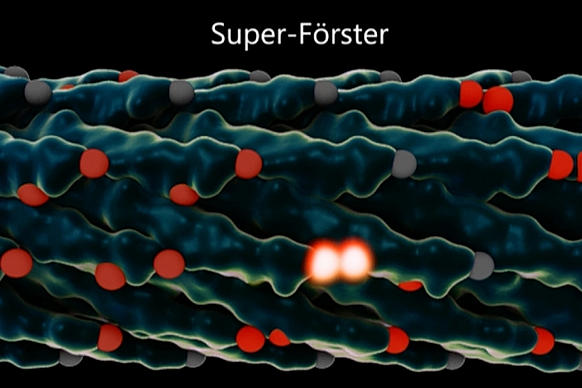
Lloyd, a professor of mechanical engineering, explains that in photosynthesis, a photon hits a receptor called a chromophore, which in turn produces an exciton — a quantum particle of energy. This exciton jumps from one chromophore to another until it reaches a reaction center, where that energy is harnessed to build the molecules that support life.

But the hopping pathway is random and inefficient unless it takes advantage of quantum effects that allow it, in effect, to take multiple pathways at once and select the best ones, behaving more like a wave than a particle.


This efficient movement of excitons has one key requirement: The chromophores have to be arranged just right, with exactly the right amount of space between them. This, Lloyd explains, is known as the “Quantum Goldilocks Effect.”

That’s where the virus comes in. By engineering a virus that Belcher has worked with for years, the team was able to get it to bond with multiple synthetic chromophores — or, in this case, organic dyes. The researchers were then able to produce many varieties of the virus, with slightly different spacings between those synthetic chromophores, and select the ones that performed best.

In the end, they were able to more than double excitons’ speed, increasing the distance they traveled before dissipating — a significant improvement in the efficiency of the process.

The project started from a chance meeting at a conference in Italy. Lloyd and Belcher, a professor of biological engineering, were reporting on different projects they had worked on, and began discussing the possibility of a project encompassing their very different expertise. Lloyd, whose work is mostly theoretical, pointed out that the viruses Belcher works with have the right length scales to potentially support quantum effects.

In 2008, Lloyd had published a paper demonstrating that photosynthetic organisms transmit light energy efficiently because of these quantum effects. When he saw Belcher’s report on her work with engineered viruses, he wondered if that might provide a way to artificially induce a similar effect, in an effort to approach nature’s efficiency.

“I had been talking about potential systems you could use to demonstrate this effect, and Angela said, ‘We’re already making those,’” Lloyd recalls. Eventually, after much analysis, “We came up with design principles to redesign how the virus is capturing light, and get it to this quantum regime.”

Within two weeks, Belcher’s team had created their first test version of the engineered virus. Many months of work then went into perfecting the receptors and the spacings.

Once the team engineered the viruses, they were able to use laser spectroscopy and dynamical modeling to watch the light-harvesting process in action, and to demonstrate that the new viruses were indeed making use of quantum coherence to enhance the transport of excitons.

“It was really fun,” Belcher says. “A group of us who spoke different [scientific] languages worked closely together, to both make this class of organisms, and analyze the data. That’s why I’m so excited by this.”

While this initial result is essentially a proof of concept rather than a practical system, it points the way toward an approach that could lead to inexpensive and efficient solar cells or light-driven catalysis, the team says. So far, the engineered viruses collect and transport energy from incoming light, but do not yet harness it to produce power (as in solar cells) or molecules (as in photosynthesis). But this could be done by adding a reaction center, where such processing takes place, to the end of the virus where the excitons end up.

“This is exciting and high-quality research,” says Alán Aspuru-Guzik, a professor of chemistry and chemical biology at Harvard University who was not involved in this work. The research, he says, “combines the work of a leader in theory (Lloyd) and a leader in experiment (Belcher) in a truly multidisciplinary and exciting combination that spans biology to physics to potentially, future technology.”

“​Access to controllable excitonic systems is a goal shared by many researchers in the field,” Aspuru-Guzik adds. “This work provides fundamental understanding that can allow for the development of devices with an increased control of exciton flow.”

The research was supported by the Italian energy company Eni through the MIT Energy Initiative. In addition to MIT postdocs Nimrod Heldman and Patrick Rebentrost, the team included researchers at the University of Florence, the University of Perugia, and Eni.

Tuesday, June 9, 2015

MIPT Physicists Develop Ultrasensitive Nanomechanical Biosensor


Two young researchers working at the MIPT Laboratory of Nanooptics and Plasmonics, Dmitry Fedyanin and Yury Stebunov, have developed an ultracompact highly sensitive nanomechanical sensor for analyzing the chemical composition of substances and detecting biological objects, such as viral disease markers, which appear when the immune system responds to incurable or hard-to-cure diseases, including HIV, hepatitis, herpes, and many others. The sensor will enable doctors to identify tumor markers, whose presence in the body signals the emergence and growth of cancerous tumors.

The sensitivity of the new device is best characterized by one key feature: according to its developers, the sensor can track changes of just a few kilodaltons in the mass of a cantilever in real time. One Dalton is roughly the mass of a proton or neutron, and several thousand Daltons are the mass of individual proteins and DNA molecules. So the new optical sensor will allow for diagnosing diseases long before they can be detected by any other method, which will pave the way for a new-generation of diagnostics.

The device, described in an article published in the journal Scientific Reports, is an optical or, more precisely, optomechanical chip. “We’ve been following the progress made in the development of micro- and nanomechanical biosensors for quite a while now and can say that no one has been able to introduce a simple and scalable technology for parallel monitoring that would be ready to use outside a laboratory. So our goal was not only to achieve the high sensitivity of the sensor and make it compact, but also make it scalabile and compatibile with standard microelectronics technologies,” the researchers said.

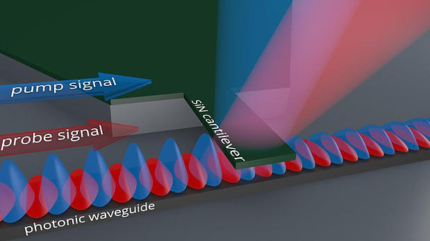
Unlike similar devices, the new sensor has no complex junctions and can be produced through a standard CMOS process technology used in microelectronics. The sensor doesn’t have a single circuit, and its design is very simple. It consists of two parts: a photonic (or plasmonic) nanowave guide to control the optical signal, and a cantilever hanging over the waveguide.

A cantilever, or beam, is a long and thin strip of microscopic dimensions (5 micrometers long, 1 micrometer wide and 90 nanometers thick), connected tightly to a chip. To get an idea how it works, imagine you press one end of a ruler tightly to the edge of a table and allow the other end to hang freely in the air. If you touch the latter with your other hand and then take your hand away, the ruler will start making mechanical oscillations at a certain frequency. That’s how the cantilever works. The difference between the oscillations of the ruler and the cantilever is only the frequency, which depends on the materials and geometry: while the ruler oscillates at several tens of hertz, the frequency of the cantilever’s oscillations is measured in megahertz. In other words, it makes a few million oscillations per second!


There are two optical signals going through the waveguide during oscillations: the first one sets the cantilever in motion, and the second one allows for reading the signal containing information about the movement. The inhomogeneous electromagnetic field of the control signal’s optical mode transmits a dipole moment to the cantilever, impacting the dipole at the same time so that the cantilever starts to oscillate. 

The sinusoidally modulated control signal makes the cantilever oscillate at an amplitude of up to 20 nanometers. The oscillations determine the parameters of the second signal, the output power of which depends on the cantilever’s position.


The highly localized optical modes of nanowave guides, which create a strong electric field intensity gradient, are key to inducing cantilever oscillations. Because the changes of the electromagnetic field in such systems are measured in tens of nanometers, researchers use the term “nanophotonics” – so the prefix “nano” is not used here just as a fad! Without the nanoscale waveguide and the cantilever, the chip simply wouldn’t work. Abig cantilever cannot be made to oscillate by freely propagating light, and the effects of chemical changes to its surface on the oscillation frequency would be less noticeable..

Cantilever oscillations make it possible to determine the chemical composition of the environment in which the chip is placed. That’s because the frequency of mechanical vibrations depends not only on the materials’ dimensions and properties, but also on the mass of the oscillatory system, which changes during a chemical reaction between the cantilever and the environment. By placing different reagents on the cantilever, researchers make it react with specific substances or even biological objects. If you place antibodies to certain viruses on the cantilever, it’ll capture the viral particles in the analyzed environment. Oscillations will occur at a lower or higher amplitude depending on the virus or the layer of chemically reactive substances on the cantilever, and the electromagnetic wave passing through the waveguide will be dispersed by the cantilever differently, which can be seen in the changes of the intensity of the readout signal.

Calculations done by the researchers showed that the new sensor will combine high sensitivity with a comparative ease of production and miniature dimensions, allowing it to be used in all portable devices, such as smartphones, wearable electronics, etc. One chip, several millimeters in size, will be able to accommodate several thousand such sensors, configured to detect different particles or molecules. The price, thanks to the simplicity of the design, will most likely depend on the number of sensors, being much more affordable than its competitors.

This work was financed by the Russian Ministry of Education and Science through state order # 16.19.2014 / K. 

Source: http://www.nanotechnologyworld.org/#!MIPT-Physicists-Develop-Ultrasensitive-Nanomechanical-Biosensor/c89r/55770f270cf293eac8071cb0