Showing posts with label DNA. Show all posts

Thursday, June 23, 2016

Making error-free DNA from RNA


For 3 billion years, one of the major carriers of information needed for life, RNA, has had a glitch that creates errors when making copies of genetic information. Researchers at The University of Texas at Austin have developed a fix that allows RNA to accurately proofread for the first time. The new discovery, published June 23 in the journal Science, will increase precision in genetic research and could dramatically improve medicine based on a person's genetic makeup.

Certain viruses called retroviruses can cause RNA to make copies of DNA, a process called reverse transcription. This process is notoriously prone to errors because an evolutionary ancestor of all viruses never had the ability to accurately copy genetic material.

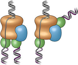
The new innovation engineered at UT Austin is an enzyme that performs reverse transcription but can also "proofread," or check its work while copying genetic code. The enzyme allows, for the first time, for large amounts of RNA information to be copied with near perfect accuracy.

"We created a new group of enzymes that can read the genetic information inside living cells with unprecedented accuracy," says Jared Ellefson, a postdoctoral fellow in UT Austin's Center for Systems and Synthetic Biology. "Overlooked by evolution, our enzyme can correct errors while copying RNA."

Reverse transcription is mainly associated with retroviruses such as HIV. In nature, these viruses' inability to copy DNA accurately may have helped create variety in species over time, contributing to the complexity of life as we know it.

Since discovering reverse transcription, scientists have used it to better understand genetic information related to inheritable diseases and other aspects of human health. Still, the error-prone nature of existing RNA sequencing is a problem for scientists.

"With proofreading, our new enzyme increases precision and fidelity of RNA sequencing," says Ellefson. "Without the ability to faithfully read RNA, we cannot accurately determine the inner workings of cells. These errors can lead to misleading data in the research lab and potential misdiagnosis in the clinical lab."

Ellefson and the team of researchers engineered the new enzyme using directed evolution to train a high-fidelity (proofreading) DNA polymerase to use RNA templates. The new enzyme, called RTX, retains the highly accurate and efficient proofreading function, while copying RNA. Accuracy is improved at least threefold, and it may be up to 10 times as accurate. This new enzyme could enhance the methods used to read RNA from cells.

"As we move towards an age of personalized medicine where everyone's transcripts will be read out almost as easily as taking a pulse, the accuracy of the sequence information will become increasingly important," said Andy Ellington, a professor of molecular biosciences. "The significance of this is that we can now also copy large amounts of RNA information found in modern genomes, in the form of the RNA transcripts that encode almost every aspect of our physiology. This means that diagnoses made based on genomic information are far more likely to be accurate. "

Reference:

Synthetic evolutionary origin of a proofreading reverse transcriptase
Science  24 Jun 2016:
Vol. 352, Issue 6293, pp. 1590-1593
DOI: 10.1126/science.aaf5409


Nanotechnology World Association


Monday, November 2, 2015

First Complete Pictures of Cells' DNA-Copying Machinery


The first-ever images of the protein complex that unwinds, splits, and copies double-stranded DNA reveal something rather different from the standard textbook view. The electron microscope images, created by scientists at the U.S. Department of Energy's Brookhaven National Laboratory with partners from Stony Brook University and Rockefeller University, offer new insight into how this molecular machinery functions, including new possibilities about its role in DNA "quality control" and cell differentiation. The images and implications are described in a paper published online by the journal Nature Structural & Molecular Biology, November 2, 2015.

"This work is a continuation of our long-standing research using electron microscopy to understand the mechanism of DNA replication, an essential function for every living cell," said Huilin Li, a biologist with a joint appointment at Brookhaven Lab and Stony Brook University.

"These new images show the fully assembled and fully activated 'helicase' protein complex—which encircles and separates the two strands of the DNA double helix as it passes through a central pore in the structure—and how the helicase coordinates with the two 'polymerase' enzymes that duplicate each strand to copy the genome."

Three-dimensional structure of the active DNA helicase bound to the front-end DNA polymerase (Pol epsilon). The DNA polymerase epsilon (green) sits on top rather than the bottom of the helicase.


Studying this molecular machinery, known collectively as a "replisome," and the details of its DNA-copying process can help scientists understand what happens when DNA is miscopied—a major source of mutation that can lead to cancer—or learn more about how a single cell can eventually develop into the many cell types that make up a multicellular organism. But no one has produced a real structure of a replisome at any resolution for any organism until now.

"All the textbook drawings and descriptions of how a replisome should look and work are based on biochemical and genetic studies," Li said, likening the situation to the famous parable of the three blind men trying to describe an elephant, each looking at only one part. Those textbook drawings show the helicase moving along the DNA, separating the two strands of the double helix, with two polymerases located at the back where the DNA strand is split. In this configuration, the polymerases would add nucleotides (molecules containing the complementary A, T, G, and C bases of the genetic code) to the side-by-side split ends as they move out of the helicase to form two new complete double helix DNA strands.

Collaborating scientists and study coauthors Zuanning Yuan, a graduate student at Stony Brook University (standing), Huilin Li of Stony Brook and Brookhaven Lab (seated, back), and Jingchuan Sun of Brookhaven Lab (seated, front) examining protein structures.

To test these assumptions, Li's group turned to the technique they had previously used to study individual components of the helicase, electron microscopy (EM). Jingchuan Sun, an EM expert in Li's lab, was essential to the success of the work. He studied samples of replisomes from baker's yeast cells—a model for all nucleus-containing cells—prepared and provided by Roxana Georgescu in Michael O'Donnell's research group at Rockefeller University. O'Donnell's group had previously published biochemical results related to this work.

"DNA replication is one of the most fundamental processes of life, so it is every biochemist's dream to see what a replisome looks like," Sun said. "Our lab has expertise and a decade of experience using electron microscopy to study DNA replication, which has prepared us well to tackle the highly mobile therefore very challenging replisome structure. Working together with the O'Donnell lab, which has done beautiful functional studies on the yeast replisome, our two groups brought perfectly complementary expertise to this project," he said.

The team's first-ever images of an intact replisome revealed that only one of the polymerases is located at the back of the helicase. The other is on the front side of the helicase, where the helicase first encounters the double-stranded helix. This means that while one of the two split DNA strands is acted on by the polymerase at the back end, the other has to thread itself back through or around the helicase to reach the front-side polymerase before having its new complementary strand assembled.

The scientists were so surprised by this finding that they asked another group at Rockefeller, led by Brian Chait, to perform additional structural studies using mass spectrometry. Yi Shi, a postdoctoral fellow in Chait's group performed this work, which confirmed the electron-microscopy-based conclusion about the unexpected architecture of the replisome.

The counterintuitive position of one polymerase at the front of the helicase suggests that it may have an unforeseen function. The authors suggest several possibilities including keeping the two "daughter" strands separate to help organize them during replication and cell division. It might also be possible that, as the single strand moves over other portions of the structure, some "surveillance" protein components check for lesions or mistakes in the nucleotide sequence before it gets copied—a sort of molecular quality control.

This architecture could also potentially play an important role in developmental biology by providing a pathway for treating the two daughter strands differently. Many modifications to DNA, including how it is packaged with other proteins, control which of the many genes in the sequence are eventually expressed in cells. An asymmetric replisome may result in asymmetric treatment of the two daughter strands during cell division—an essential step for making different tissues within a multicellular organism.

As the paper concludes, "Clearly, further studies will be required to understand the functional implications of the unexpected replisome architecture reported here."

This research was funded by the National Institutes of Health (GM103314, GM109824, GM74985, AG29979, and GM38839) and by the Howard Hughes Medical Institute.

Wednesday, October 7, 2015

Detecting HIV diagnostic antibodies with DNA nanomachines


A nanoscale machine composed of synthetic DNA can be used for the rapid, sensitive and low-cost diagnosis of many diseases, including HIV

New research may revolutionize the slow, cumbersome and expensive process of detecting the antibodies that can help with the diagnosis of infectious and auto-immune diseases such as rheumatoid arthritis and HIV. An international team of researchers have designed and synthetized a nanometer-scale DNA "machine" whose customized modifications enable it to recognize a specific target antibody. Their new approach, which they described this month in Angewandte Chemie, promises to support the development of rapid, low-cost antibody detection at the point-of-care, eliminating the treatment initiation delays and increasing healthcare costs associated with current techniques.

The binding of the antibody to the DNA machine causes a structural change (or switch), which generates a light signal. The sensor does not need to be chemically activated and is rapid - acting within five minutes - enabling the targeted antibodies to be easily detected, even in complex clinical samples such as blood serum.

"One of the advantages of our approach is that it is highly versatile," said Prof. Francesco Ricci, of the University of Rome, Tor Vergata, senior co-author of the study. "This DNA nanomachine can be in fact custom-modified so that it can detect a huge range of antibodies, this makes our platform adaptable for many different diseases".

"Our modular platform provides significant advantages over existing methods for the detection of antibodies," added Prof. Vallée-Bélisle of the University of Montreal, the other senior co-author of the paper. "It is rapid, does not require reagent chemicals, and may prove to be useful in a range of different applications such as point-of-care diagnostics and bioimaging".

"Another nice feature of our this platform is its low-cost," said Prof. Kevin Plaxco of the University of California, Santa Barbara. "The materials needed for one assay cost about 15 cents, making our approach very competitive in comparison with other quantitative approaches."

"We are excited by these preliminary results, but we are looking forward to improve our sensing platform even more" said Simona Ranallo, a PhD student in the group of Prof. Ricci at the University of Rome and first-author of the paper. "For example, we could adapt our platform so that the signal of the nanoswitch may be read using a mobile phone. This will make our approach really available to anyone! We are working on this idea and we would like to start involving diagnostic companies."


Thursday, September 24, 2015

Tracking nanowalkers with light

A gold cylinder with DNA feet can climb over DNA-primed hills made from folded DNA strands. The second cylinder (red) serves as a point of reference for observing the nanowalker. © MPI for Intelligent Systems, Stuttgart

A tiny gold rod walks across a surface guided by DNA and can be tracked step by step

Nanotechnology is taking its first steps. Researchers from the Max Planck Institute for Intelligent Systems in Stuttgart have developed a gold nanocylinder equipped with discrete DNA strands as ‘feet’ that can walk across a DNA origami platform. They are able to trace the movements of the nanowalker, which is smaller than the optical resolution limit, by exciting plasmons in the gold nanocylinder. Plasmons are collective oscillations of numerous electrons. The excitation changes the ray of light, thus allowing the researchers to actually observe the nanowalker. Their main objective is to use such mobile plasmonic nanoobjects to study how miniscule particles interact with light.

The body of the nanowalker consists of a gold cylinder that is 35 nanometres long and ten nanometres wide. “The cylinder’s surface is primed with numerous identical strands of DNA that effectively serve as feet,” Group Leader Liu explains. These DNA strands stick out from the gold cylinder like the bristles of a bottle brush. “They allow the gold cylinder to make contact with the surface underneath and travel across it.”Nanomachines – i.e. mechanical devices with dimensions of nanometers – could one day carry out specific tasks in fields such as medicine, information processing, chemistry or scientific research, according to nanotechnology experts. Yet miniature machines that are thousands of times smaller than the diameter of a human hair pose significant challenges for scientists: firstly, the individual constituents merely consist of a small number of atoms; it is barely possible to handle such components, let alone assemble them in a precise manner. Moreover, the machines would then need to be supplied with energy. And ultimately, the researchers cannot simply check to see if their device is in fact working. The microscopy techniques necessary for such observation are complex and require for example vacuum chambers, in which the devices would be destroyed. At the Max Planck Institute for Intelligent Systems in Stuttgart, a team of researchers including Chao Zhou and Xiaoyang Duan, headed by Laura Na Liu has now created a nanowalker that they can observe with the help of a nanooptical effect.

The nanowalker strides across a carpet of DNA strands

The gold cylinder’s walkway is composed of DNA as well – a DNA origami template, to be precise. Extended from this folded DNA scaffold like fibres from a carpet are longitudinal rows of short strands that are parallel to the cylinder and serve as footholds for the walker’s tiny feet. Each row in the DNA carpet comprises a different combination of bases, and each row represents one station. Initially, the walker’s feet bind with two neighbouring rows, while the footholds of the other rows remain blocked.
“The walker moves forward in a rolling motion, from station to station,” says Liu. In order to make this possible, the researchers must constantly add short snippets of DNA to the fluid in which the action is taking place. These snippets are designed to match the DNA of the individual rows. First they break up a row of connections linking the walker’s feet and the DNA of the platform and block the footholds of that particular station. On the opposite side of the walker, they then unblock a separate row, to which the cylinder’s feet can now attach.
“Depending on what is added, the walker moves either in one direction or in the other,” explains Liu. “We are inspired by naturally occurring molecular motors: The fluid moves the cylinder and its feet back and forth by means of thermal motion.” Due to the fact that the feet only ever redock on one side, the walker slowly moves forward. Each step is seven nanometres long, which is over one hundred thousand times smaller than the single stride of a wood ant.

Researchers use plasmon resonance to trace the nanocylinder’s path

In order to trace the tiny machine’s path, the researchers relied on a nanooptical effect called plasmon resonance. Plasmons are collective oscillations of numerous electrons and are often present in metals, among other materials. “Light can interact with the plasmons in the gold,” Liu explains. “Light is partially absorbed in the process in our case, resulting in what is known as plasmon resonance.” By analysing the light beam, the researchers can measure this phenomenon.
Determining the cylinder’s exact location, however, required placing a second, stationary gold nanocylinder on the underside of the DNA origami platform. Broadly speaking, this second cylinder serves as a point of reference. The reason for this is because together, the two cylinders bring about a change in the circular polarisation of the light beam: Light consists of an oscillating electromagnetic field. The polarisation is equivalent to the direction in which the field oscillates; in circularly polarised light, it turns either clockwise or counterclockwise. By observing the spectral changes resulting from the interaction with circular polarized light, the researchers can determine the walker’s current position.
“By using this approach we were able to trace every single step. That’s why the walker is more than just a mobile element – it also provides information about its location,” says Liu. Sophisticated microscope technology thus became redundant for observing the plasmonic walker, which Liu deems a precursor of a “new generation of nanomachines with customised optical properties”. The researcher now aims to use this tool to further study the interaction of light and matter on a nanoscale, as well as the mechanical behaviour of nanoparticles. Because if the gold walker is indeed destined to one day reach its goal and complete various tasks, it still needs to take quite a few strides – and not just on DNA origami.

Read more on Nanotechnology World Association

Thursday, September 3, 2015

Using DNA origami to build nanodevices of the future


Scientists have been studying ways to use synthetic DNA as a building block for smaller and faster devices. DNA has the advantage of being inherently "coded". Each DNA strand is formed of one of four "codes" that can link to only one complementary code each, thus binding two DNA strands together. Scientists are using this inherent coding to manipulate and "fold" DNA to form "origami nanostructures": extremely small two- and three-dimensional shapes that can then be used as construction material to build nanodevices such as nanomotors for use in targeted drug delivery inside the body.

Despite progress that has been made in this field, assembling DNA origami units into larger structures remains challenging.

A team of scientists at Kyoto University's Institute for Integrated Cell-Material Sciences (iCeMS) has developed an approach that could bring us one step closer to the nanomachines of the future.

They used a double layer of lipids (fats) containing both a positive and a negative charge. DNA origami structures were weakly absorbed onto the lipid layer through an electrostatic interaction.

The weak bond between the origami structures and the lipid layer allowed them to move more freely than in other approaches developed by scientists, facilitating their interaction with one another to assemble and form larger structures.

"We anticipate that our approach will further expand the potential applications of DNA origami structures and their assemblies in the fields of nanotechnology, biophysics and synthetic biology," says chemical biologist Professor Hiroshi Sugiyama from iCeMS.

http://www.nanotechnologyworld.org/#!Using-DNA-origami-to-build-nanodevices-of-the-future/c89r/55e5c5cb0cf2c1d1fd664f9b

Monday, June 8, 2015

UCLA researchers discover molecular rules that govern autoimmune disorders


An international team led by researchers at UCLA’s Henry Samueli School of Engineering and Applied Science and California NanoSystems Institute has identified an unexpectedly general set of rules that determine which molecules can cause the immune system to become vulnerable to the autoimmune disorders lupus and psoriasis.
The breakthrough, described in an article published today in the journal Nature Materials, could lead to new ways of treating the disorders.

Led by Gerard C. L. Wong, a UCLA professor of bioengineering and chemistry who is affiliated with CNSI, the multidisciplinary team also included Michel Gilliet of Switzerland’s Lausanne University Hospital, and Jure Dobnikar and Daan Frenkel of the University of Cambridge. 

Autoimmune diseases strike when the body attacks itself because it fails to distinguish between host tissue and disease-causing agents, or pathogens. Two such disorders are lupus, which can damage the skin, joints and organs, causing rashes, hair loss and fatigue; and psoriasis, which causes rashes, lesions and arthritis, and creates an increased risk for cancer and diabetes.

When a healthy person is infected by a virus, viral DNA can activate immune cells via a receptor called TLR9. The receptor triggers the cells to send signaling molecules called interferons to initiate a powerful defensive response.
In people with lupus or psoriasis, these cells are activated by their own DNA, or self-DNA. 

Using synchrotron X-ray scattering and other techniques, researchers determined that a broad range of molecules, both organic and inorganic, can organize self-DNA into a liquid crystalline structure that binds strongly to the TLR9 receptors — like the teeth on either side of a zipper. This structure protects the DNA from becoming degraded and greatly amplifies the body’s immune response.

Synchrotron X-ray scattering utilizes a particle accelerator to generate X-ray beams that allow researchers to determine how atoms and molecules are organized into different structures.

“Our research has identified a set of rules that tell us what types of molecules or materials can set off this aspect of the immune system,” Wong said. “This new knowledge will make it easier to design new therapeutic strategies to control immune responses.”

Nathan Schmidt, one of the paper’s lead authors, said, “Our colleagues had established empirically that certain molecules were activating self-DNA and triggering responses in disorders such as lupus and psoriasis. We were able to elucidate something that was poorly understood — a key to triggering the immune response is that the molecules must arrange the DNA so that the receptors bind to them strongly.”

Schmidt, a former member of Wong’s lab, is now a postdoctoral researcher at UC San Francisco. Fan Jin, another lead author, was a member of Wong’s lab at UCLA and is now a professor at the University of Science and Technology of China. Other authors include postdoctoral researcher Wujing Xian and graduate student researcher Calvin Lee, both of Wong’s lab, as well as researchers in England, Italy, Slovenia and Switzerland.

The research was supported by the National Institutes of Health, the National Science Foundation, the European Commission, the European Research Council, the Slovenian Research Agency, the Swiss National Science Foundation and the University of Cambridge.

Source: http://www.nanotechnologyworld.org/#!UCLA-researchers-discover-molecular-rules-that-govern-autoimmune-disorders/c89r/55760e560cf2e4994fb916a3

Monday, May 25, 2015

DNA Double Helix Does Double Duty in Assembling Arrays of Nanoparticles


octahedral frames
A combination cryo-electron microscopy image of an octahedral frame with one gold nanoparticle bound to each of the six vertices, shown from three different angles
Synthetic pieces of biological molecule form framework and glue for making nanoparticle clusters and arrays

In a new twist on the use of DNA in nanoscale construction, scientists at the U.S. Department of Energy's (DOE) Brookhaven National Laboratory and collaborators put synthetic strands of the biological material to work in two ways: They used ropelike configurations of the DNA double helix to form a rigid geometrical framework, and added dangling pieces of single-stranded DNA to glue nanoparticles in place. 
The method, described in the journal Nature Nanotechnology, produced predictable clusters and arrays of nanoparticles—an important step toward the design of materials with tailored structures and functions for applications in energy, optics, and medicine.
"These arrays of nanoparticles with predictable geometric configurations are somewhat analogous to molecules made of atoms," said Brookhaven physicist Oleg Gang, who led the project at the Lab's Center for Functional Nanomaterials (CFN), a DOE Office of Science User Facility. "While atoms form molecules based on the nature of their chemical bonds, there has been no easy way to impose such a specific spatial binding scheme on nanoparticles. This is exactly the problem that our method addresses." 
Using the new method, the scientists say they can potentially orchestrate the arrangements of different types of nanoparticles to take advantage of collective or synergistic effects. Examples could include materials that regulate energy flow, rotate light, or deliver biomolecules. 
"We may be able to design materials that mimic nature's machinery to harvest solar energy, or manipulate light for telecommunications applications, or design novel catalysts for speeding up a variety of chemical reactions," Gang said.
octahedrons
Scientists built octahedrons using ropelike structures made of bundles 
of DNA double-helix molecules to form the frames (a). Single strands of 
DNA attached at the vertices (numbered in red) can be used to attach 
nanoparticles coated with complementary strands. This approach can 
yield a variety of structures, including ones with the same type of particle 
at each vertex (b), arrangements with particles placed only on certain 
vertices (c), and structures with different particles placed strategically 
on different vertices (d).
"We may be able to design materials that harvest solar energy, manipulate light, or speed up a variety of chemical reactions." — Brookhaven physicist Oleg Gang

The scientists demonstrated the technique to engineer nanoparticle architectures using an octahedral scaffold with particles positioned in precise locations on the scaffold according to the specificity of DNA coding. The designs included two different arrangements of the same set of particles, where each configuration had different optical characteristics. They also used the geometrical clusters as building blocks for larger arrays, including linear chains and two-dimensional planar sheets.
"Our work demonstrates the versatility of this approach and opens up numerous exciting opportunities for high-yield precision assembly of tailored 3D building blocks in which multiple nanoparticles of different structures and functions can be integrated," said CFN scientist Ye Tian, one of the lead authors on the paper.
Details of assembly
This nanoscale construction approach takes advantage of two key characteristics of the DNA molecule: the twisted-ladder double helix shape, and the natural tendency of strands with complementary bases (the A, T, G, and C letters of the genetic code) to pair up in a precise way. 
First, the scientists created bundles of six double-helix molecules, then put four of these bundles together to make a stable, somewhat rigid building material—similar to the way individual fibrous strands are woven together to make a very strong rope. The scientists then used these ropelike girders to form the frame of three-dimensional octahedrons, "stapling" the linear DNA chains together with hundreds of short complementary DNA strands.
"We refer to these as DNA origami octahedrons," Gang said.
To make it possible to "glue" nanoparticles to the 3D frames, the scientists engineered each of the original six-helix bundles to have one helix with an extra single-stranded piece of DNA sticking out from both ends. When assembled into the 3D octahedrons, each vertex of the frame had a few of these "sticky end" tethers available for binding with objects coated with complementary DNA strands.
"When nanoparticles coated with single strand tethers are mixed with the DNA origami octahedrons, the 'free' pieces of DNA find one another so the bases can pair up according to the rules of the DNA complementarity code. Thus the specifically DNA-encoded particles can find their correspondingly designed place on the octahedron vertices" Gang said.
The scientists can change what binds to each vertex by changing the DNA sequences encoded on the tethers. In one experiment, they encoded the same sequence on all the octahedron's tethers, and attached strands with a complementary sequence to gold nanoparticles. The result: One gold nanoparticle attached to each of octahedron's six vertices. 
In additional experiments the scientists changed the sequence of some vertices and used complementary strands on different kinds of particles, illustrating that they could direct the assembly and arrangement of the particles in a very precise way. In one case they made two different arrangements of the same three pairs of particles of different sizes, producing products with different optical properties. They were even able to use DNA tethers on selected vertices to link octahedrons end to end, forming chains, and in 2D arrays, forming sheets.
Visualization of arrays
octahedrons
By strategically placing tethers on particular vertices, the scientists
used the octahedrons to link nanoparticles into one-dimensional
chainlike arrays (left) and two-dimensional square sheets (right). 
Confirming the particle arrangements and structures was a major challenge because the nanoparticles and the DNA molecules making up the frames have very different densities. Certain microscopy techniques would reveal only the particles, while others would distort the 3D structures. 
To see both the particles and origami frames, the scientists used cryo-electron microscopy (cryo-EM), led by Brookhaven Lab and Stony Brook University biologist Huilin Li, an expert in this technique, and Tong Wang, the paper's other lead co-author, who works in Brookhaven's Biosciences department with Li. They had to subtract information from the images to "see" the different density components separately, then combine the information using single particle 3D reconstruction and tomography to produce the final images. 
"Cryo-EM preserves samples in their near-native states and provides close to nanometer resolution," Wang said. "We show that cryo-EM can be successfully applied to probe the 3D structure of DNA-nanoparticle clusters."
These images confirm that this approach to direct the placement of nanoparticles on DNA-encoded vertices of molecular frames could be a successful strategy for fabricating novel nanomaterials.
This research was supported by the DOE Office of Science.
Brookhaven National Laboratory is supported by the Office of Science of the U.S. Department of Energy.  The Office of Science is the single largest supporter of basic research in the physical sciences in the United States, and is working to address some of the most pressing challenges of our time.  For more information, please visit science.energy.gov.