Showing posts with label nanoscale. Show all posts

Wednesday, June 10, 2015

Rice researchers make ultrasensitive conductivity measurements


Researchers at Rice University have discovered a new way to make ultrasensitive conductivity measurements at optical frequencies on high-speed nanoscale electronic components.

The research at Rice’s Laboratory for Nanophotonics (LANP) is described online in a new study in the American Chemical Society’s journal ACS Nano. In a series of experiments, LANP researchers linked pairs of puck-shaped metal nanodisks with metallic nanowires and showed how the flow of current at optical frequencies through the nanowires produced “charge transfer plasmons” with unique optical signatures.

“The push to continually increase the speed of microchip components has researchers looking at nanoscale devices and components that operate at optical frequencies for next-generation electronics,” said LANP Director Naomi Halas, the lead scientist on the study. “It is not well-known how these materials and components operate at extremely high frequencies of light, and LANP’s new technique provides a way to measure the electrical transport properties of nanomaterials and structures at these extremely high frequencies.”

Read more: http://www.nanotechnologyworld.org/#!Rice-researchers-make-ultrasensitive-conductivity-measurements/c89r/5578553e0cf2df2eae412b28 

Monday, May 26, 2014

Fractal Nanotruss Work


Fancy Erector Set? Nope. The elaborate fractal structure shown at right is many, many times smaller than that and is certainly not child's play. It is the latest example of what Julia Greer, professor of materials science and mechanics, calls a fractal nanotruss—nano because the structures are made up of members that are as thin as five nanometers (five billionths of a meter); truss because they are carefully architected structures that might one day be used in structural engineering materials.


Greer's group has developed a three-step process for building such complex structures very precisely. They first use a direct laser writing method called two-photon lithography to "write" a three-dimensional pattern in a polymer, allowing a laser beam to crosslink and harden the polymer wherever it is focused. At the end of the patterning step, the parts of the polymer that were exposed to the laser remain intact while the rest is dissolved away, revealing a three-dimensional scaffold. Next, the scientists coat the polymer scaffold with a continuous, very thin layer of a material—it can be a ceramic, metal, metallic glass, semiconductor, "just about anything," Greer says. In this case, they used alumina, or aluminum oxide, which is a brittle ceramic, to coat the scaffold. In the final step they etch out the polymer from within the structure, leaving a hollow architecture.

Taking advantage of some of the size effects that many materials display at the nanoscale, these nanotrusses can have unusual, desirable qualities. For example, intrinsically brittle materials, like ceramics, including the alumina shown, can be made deformable so that they can be crushed and still rebound to their original state without global failure.

"Having full control over the architecture gives us the ability to tune material properties to what was previously unattainable with conventional monolithic materials or with foams," says Greer. "For example, we can decouple strength from density and make materials that are both strong (and tough) as well as extremely lightweight. These structures can contain nearly 99 percent air yet can also be as strong as steel. Designing them into fractals allows us to incorporate hierarchical design into material architecture, which promises to have further beneficial properties."

The members of Greer's group who helped develop the new fabrication process and created these nanotrusses are graduate students Lucas Meza and Lauren Montemayor and Nigel Clarke, an undergraduate intern from the University of Waterloo.
Written by Kimm Fesenmaier

Source: http://www.caltech.edu/content/miniature-truss-work#sthash.xl98f5Tu.dpuf

Thursday, May 22, 2014

Resonant Energy Transfer from Quantum Dots to Graphene

Schematic of a quantum dot-graphene nano-photonic device,
as described in this research project.
Semiconductor quantum dots (QDs) are nanoscale semiconductors that exhibit size dependent physical properties. For example, the color (wavelength) of light that they absorb changes dramatically as the diameter decreases. 
Graphene is an atomically thick sheet of carbon atoms, arranged in a hexagonal lattice pattern. In this work, QDs have been combined with graphene to develop nanoscale photonic devices that can dramatically improve our ability to detect light. Quantum dots can absorb light and transfer it to graphene, but the efficiency of the transfer depends on how far the QDs and the graphene are separated from each other. 
This study demonstrated that the thickness of the organic molecule layer that typically surrounds the QDs is crucial in attaining sufficiently high efficiency of this light/energy transfer into the graphene. In other works, the thinner the organic layer, the better. This transfer can be further optimized by engineering the interface between the two nanomaterials, specifically optimizing the thickness of the organic capping molecules on the quantum dots. Based on this work, further improvement of the performance of these nano-photonic devices can be expected.

Why Does This Matter?

chloride-terminated CdSe quantum dot
a) Schematic of a chloride-terminated CdSe quantum dot. b) A high resolution transmission electron microscopy image of such quantum dots.
Commercial cadmium selenide (CdSe) quantum dots have long insulating organic ligands that prevent their utilization in energy and charge transfer applications for which short distances between the QDs and other materials are critical.  Short, chlorine ligands that passivated CdSe QDs are an intriguing alternative material to enhance the interaction with materials into which charge carriers, such as electrons, can easily conduct.  Graphene is such a material.  The combination of CdSe quantum dots and graphene could hold the key to the development and implementation of nanoscale materials systems in flexible electronics and photodetectors.
Photoluminescence lifetime decay of isolated quantum dots
Photoluminescence lifetime decay of isolated quantum dots on glass (blue) and graphene (red) demonstrate efficient energy transfer between the quantum dots and graphene.

What Are The Details?

  • CFN Capabilities: The Advanced Optical Microscopy Facility measured the time-resolved photoluminescence from isolated CdSe quantum dots deposited on graphene.
  • The team discovered that short, chloride-capped CdSe quantum dots, deposited on chemical-vapor-deposited, monolayer layer graphene, exhibited highly efficient energy transfer to the graphene with a 4x observed reduction in the excitonic lifetime.  This demonstrated significant near-field coupling between quantum dots and the graphene.  

http://www.bnl.gov/newsroom/news.php?a=24906

Monday, March 31, 2014

Never say never in the nano-world

Artistic impressions of the nanoparticle in a laser trap.
(Image credits: Iñaki Gonzalez and Jan Gieseler)
Objects with sizes in the nanometer range, such as the molecular building blocks of living cells or nanotechnological devices, are continuously exposed to random collisions with surrounding molecules. In such fluctuating environments the fundamental laws of thermodynamics that govern our macroscopic world need to be rewritten. 

An international team of researchers from Barcelona, Zurich and Vienna found that a nanoparticle trapped with laser light temporarily violates the famous second law of thermodynamics, something that is impossible on human time and length scale. They report about their results in the latest issue of the prestigious scientific journal Nature Nanotechnology.
Surprises at the nanoscale

Watching a movie played in reverse often makes us laugh because unexpected and mysterious things seem to happen: glass shards lying on the floor slowly start to move towards each other, magically assemble and suddenly an intact glass jumps on the table where it gently gets to a halt. Or snow starts to from a water puddle in the sun, steadily growing until an entire snowman appears as if molded by an invisible hand. When we see such scenes, we immediately realize that according to our everyday experience something is out of the ordinary. Indeed, there are many processes in nature that can never be reversed. The physical law that captures this behavior is the celebrated second law of thermodynamics, which posits that the entropy of a system – a measure for the disorder of a system – never decreases spontaneously, thus favoring disorder (high entropy) over order (low entropy).

However, when we zoom into the microscopic world of atoms and molecules, this law softens up and looses its absolute strictness. Indeed, at the nanoscale the second law can be fleetingly violated. On rare occasions, one may observe events that never happen on the macroscopic scale such as, for example heat transfer from cold to hot which is unheard of in our daily lives. Although on average the second law of thermodynamics remains valid even in nanoscale systems, scientists are intrigued by these rare events and are investigating the meaning of irreversibility at the nanoscale.

Nanoparticles in laser traps

Recently, a team of physicists of the University of Vienna, the Institute of Photonic Sciences in Barcelona and the Swiss Federal Institute of Technology in Zürich succeeded in accurately predicting the likelihood of events transiently violating the second law of thermodynamics. They immediately put the mathematical fluctuation theorem they derived to the test using a tiny glass sphere with a diameter of less than 100 nm levitated in a trap of laser light. Their experimental set-up allowed the research team to capture the nano-sphere and hold it in place, and, furthermore, to measure its position in all three spatial directions with exquisite precision. In the trap, the nano-sphere rattles around due to collisions with surrounding gas molecules. 

By a clever manipulation of the laser trap the scientists cooled the nano-sphere below the temperature of the surrounding gas and, thereby, put it into a non-equilibrium state. They then turned off the cooling and watched the particle relaxing to the higher temperature through energy transfer from the gas molecules. The researchers observed that the tiny glass sphere sometimes, although rarely, does not behave as one would expect according to the second law: the nano-sphere effectively releases heat to the hotter surroundings rather than absorbing the heat. The theory derived by the researchers to analyze the experiment confirms the emerging picture on the limitations of the second law on the nanoscale.

Nanomachines out of equilibrium

The experimental and theoretical framework presented by the international research team in the renowned scientific journal Nature Nanotechnology has a wide range of applications. Objects with sizes in the nanometer range, such as the molecular building blocks of living cells or nanotechnological devices, are continuously exposed to a random buffeting due to the thermal motion of the molecules around them. As miniaturization proceeds to smaller and smaller scales nanomachines will experience increasingly random conditions. Further studies will be carried out to illuminate the fundamental physics of nanoscale systems out of equilibrium. The planned research will be fundamental to help us understand how nanomachines perform under these fluctuating conditions.

Original publication in Nature Nanotechnology

Dynamic Relaxation of a Levitated Nanoparticle from a Non-Equilibrium Steady State. Jan Gieseler, Romain Quidant, Christoph Dellago, and Lukas Novotny. Nature Nanotechnology AOP, February 28, 2014. DOI: 10.1038/NNANO.2014.40

Source: http://medienportal.univie.ac.at//presse/aktuelle-pressemeldungen/detailansicht/artikel/never-say-never-in-the-nano-world/

Monday, March 24, 2014

A mathematical equation that explains the behavior of nanofoams

The scientific team, made up of researchers from the Consejo Superior de Investigaciones Científicas (Spanish National Research Council) - CSIC, the Universidad Pontificia Comillas de Madrid- UPCO, and UC3M, reached this conclusion after producing and characterizing nanofoam formed by ion radiation on a silicon surface. This study, recently published in the journal, Physical Review Letters, describes the evolution of these nanostructures during the time of irradiation.

For this purpose, the scientists carried out an experiment that consisted in “bombardment” of a small silicon plate with energetic particles from a plasma. The objective was to observe how the surface of this crystal reacted to these different “attacks” from this type of ion radiation (ions are used: atoms of a gas that have lost an electron). “At the outset, we were studying other methods of erosion and looking for a rippled structure at the edge of our sample after applying this technique, but when we looked at its center we observed a cellular structure that got our attention because of its similarity to many other natural and artificial systems,” one of the authors of the study, Mario Castro, UPCO Professor, revealed.

Cellular structures that are more or less disordered can be found in many natural systems: from the hides of animals, such as a giraffe, to bath froth or beer foam, to microscopic fluid convection, basalt column landscapes or diverse crystalline materials. This particular order is also evident in artificial structures and even political ones, such as modern architecture or demarcation of provinces on maps.

“It is of interest to confirm that the same universal laws which regulate the cellular structures in other systems are also regulating at the nanoscale,” Rodolfo Cuerno from the UC3M Mathematics Department noted. “Furthermore,” he added “it is the first time that the evolution of a system of this kind is reproduced quite well by a single differential equation,” which also is applied to other systems. The validity of the model in this study means that the formation of certain self-organized patterns and the dynamics of the foam would be different manifestations of a same principle.

“The results of this study help us to understand how certain material systems evolve in the presence of an external agent, as in this case of ion radiation. In addition, there exists interest of a practical nature because of the importance of the technological applications of silicon as well as for the nanometric dimensions in which the phenomenon unfolds,” explained Luis Vázquez, from the Instituto de Ciencia de Materiales (Materials Science Institute) de Madrid at the CSIC.

The experimental observations have been carried out using an atomic force microscope, a machine with great precision. This type of microscope has enormous spatial resolution: it distinguishes variations in height up to a nanometer (the millionth part of a millimeter) and movements on a horizontal plane of up to 10 nanometers.

This research could have further future applications, since in general, methods are being sought to produce structures with nanometric dimensions for diverse uses, according to the scientists: for example, in order to obtain favorable conditions in certain catalytic chemical reactions, to optimize displacement of fluids in circuits on such small scale or in optoelectronics, to generate laser light if certain structures are sufficiently ordered.

Watch Video: https://www.youtube.com/watch?v=F2GHL0jhW84

Source: http://portal.uc3m.es/portal/page/portal/news_repository/general_news/A%20mathematical%20equation%20that%20explains%20the%20behavior%20of%20nanofo?_template=/SHARED/pl_noticias_detalle_pub_ingles

Thursday, March 20, 2014

Cold Chaos

Chaotic motion and complex chemistry might lurk at nano-Kelvin temperatures 


At sub-micro-kelvin temperatures atoms or molecules move so slowly that it is better to think of them as spread-out, wavelike things a micron or more across, many times larger than any putative bond length (typically sub-nanometer in size) that would characterize bound molecules. Experiments over several years have shown that collisions and chemical reactions do take place---surprising, considering the scant energy available to the reactants---but under the sway of wavelike, and not particle-like, considerations.
A new experiment conducted at the University of Innsbruck in Austria adds a new twist to this picture. There swarms of erbium atoms were held in a special trap and cooled to a temperature of about 300 nK. Er-166 and Er-168 are boson species, which means that these atoms can clump very closely together in a single quantum state known as Bose Einstein Condensate. But because of the erbium’s large intrinsic magnetic moment, inter-atom interactions are strong. By imposing an extra magnetic field---the better to excite Feshbach resonances, which are delicate bound molecular states---the types of collisions among atoms can be controlled. The Innsbruck physicists expected that the use of Er atoms would give rise to a number of such resonance states.
It came as a great surprise to them, therefore, to observe a hundred and more resonances rather than a dozen or so. The resonances were so great in number and so densely packed that the researchers deduce that a form of quantum chaos is at work here.
Quantum chaos, as a research subject, is only a few decades old. It features systems of particles exhibiting both quantum and chaotic behavior---chaos being usually thought of as classical-physics phenomenon. A classical system is generally deemed to be chaotic if its future course is described by nonlinear equations and if predictions of future behavior are exquisitely dependent our knowledge of the initial conditions of the system. The signature of quantum chaos is somewhat different: a dense set of energy levels with a special kind of spacing between levels.
The Innsbruck experiment represents the first instance of quantum chaos observed for ultracold atomic collisions. The results are reported in Nature in a paper published online on 12 March (1). JQI scientist Paul Julienne, as an expert on particle collisions at cold temperatures, was asked to write a commentary on this appearance of cold chaos. His essay appears in the same issue of Nature (2).

MORE THAN THEY BARGAINED FOR

As the strength of the external magnetic field is varied in the Innsbruck apparatus, new collision conditions become available. At certain field values the nearly-at-rest atoms come into resonance, and form weakly bound molecules; these molecules, when struck by a third atom, form a threefold object which can no longer be held by the trap. Thus a decrease in the atomic population marks the location of the resonance energies.
The hundred resonances actually observed while varying magnetic field across a fixed range was more than the researchers had expected and far more than had ever been observed before---typically in studies of alkali elements such as cesium. The bad news was that studying atomic collisions at these temperatures suddenly got a lot more complicated.
The good news is that all these new collision alternatives might open up new avenues in low-temperature physics research. “Part of the power and the beauty of cold atom physics,” said Julienne in his News & Views essay in Nature, “is that the scattering length can be made to take on any value by tuning a magnetic field close to a Feshbach resonance. Its value controls the two-body, few-body, and many-body physics of ultracold quantum matter. Thus, controlling the field makes the system dance to our tune.”
Still more complex than erbium atoms are molecules. They are currently hard to cool to nK temperatures because of their great complexity; energy continues to lurk in various rotational and vibrational modes of molecules. But molecular cooling is advancing and soon molecular chemistry at the coldest temperatures might be doable. It would not be surprising to find a similar kind of chaos at play there.
Source: http://jqi.umd.edu/news/cold-chaos#sthash.b1us507R.dpuf

Friday, March 14, 2014

Nanoscale optical switch breaks miniaturization barrier


An ultra-fast and ultra-small optical switch has been invented that could advance the day when photons replace electrons in the innards of consumer products ranging from cell phones to automobiles.
The new optical device can turn on and off trillions of times per second. It consists of individual switches that are only one five-hundredth the width of a human hair (200 nanometers) in diameter. This size is much smaller than the current generation of optical switches and it easily breaks one of the major technical barriers to the spread of electronic devices that detect and control light: miniaturizing the size of ultrafast optical switches.
Richard Haglund portrait
Physicist Richard Haglund has been studying the properties of vanadium dioxide for more than 20 years. (Joe Howell / Vanderbilt)
The new device was developed by a team of scientists from Vanderbilt University, University of Alabama-Birmingham, and Los Alamos National Laboratory and is described in the March 12 issue of the journal Nano Letters.
The ultrafast switch is made out of an artificial material engineered to have properties that are not found in nature. In this case, the “metamaterial” consists of nanoscale particles of vanadium dioxide (VO2) – a crystalline solid that can rapidly switch back and forth between an opaque, metallic phase and a transparent, semiconducting phase – which are deposited on a glass substrate and coated with a “nanomesh” of tiny gold nanoparticles.
The scientists report that bathing these gilded nanoparticles with brief pulses from an ultrafast laser generates hot electrons in the gold nanomesh that jump into the vanadium dioxide and cause it to undergo its phase change in a few trillionths of a second.
“We had previously triggered this transition in vanadium dioxide nanoparticles directly with lasers and we wanted to see if we could do it with electrons as well,” said Richard Haglund, Stevenson Professor of Physics at Vanderbilt, who led the study. “Not only does it work, but the injection of hot electrons from the gold nanoparticles also triggers the transformation with one fifth to one tenth as much energy input required by shining the laser directly on the bare VO2.”
Optical switch illustrations
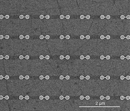
Left: Illustration of terahertz optical switches shows the vanadium dioxide nanoparticles coated with a "nanomesh" of smaller gold particles. Right: Scanning electron microscope image of the switches at two resolutions. (Haglund Lab / Vanderbilt)
Both industry and government are investing heavily in efforts to integrate optics and electronics, because it is generally considered to be the next step in the evolution of information and communications technology. Intel, Hewlett-Packard and IBM have been building chips with increasing optical functionality for the last five years that operate at gigahertz speeds, one thousandth that of the VO2 switch.
“Vanadium dioxide switches have a number of characteristics that make them ideal for optoelectronics applications,” said Haglund. In addition to their fast speed and small size, they:
  • Are completely compatible with current integrated circuit technology, both silicon-based chips and the new “high-K dielectric” materials that the semiconductor industry is developing to continue the miniaturization process that has been a major aspect of microelectronics technology development;
  • Operate in the visible and near-infrared region of the spectrum that is optimal for telecommunications applications;
  • Generate an amount of heat per operation that is low enough so that the switches can be packed tightly enough to make practical devices: about ten trillionths of a calorie (100 femtojoules) per bit.
“Vanadium dioxide’s amazing properties have been known for more than half a century. At Vanderbilt, we have been studying VO2 nanoparticles for the last ten years, but the material has been remarkably successfully at resisting theoretical explanations,” said Haglund. “It is only in the last few years that intensive computational studies have illuminated the physics that underlies its semiconductor-to-metal transition.”
Student in clean room
Graduate student Christina McGahan holding a disk on which centimeter square samples are grown. (Joe Howell / Vanderbilt)
Vanderbilt graduate students Kannatassen Appavoo and Joyeeta Nag fabricated the metamaterial at Vanderbilt; Appavoo joined forces with University of Alabama, Birmingham graduate student Nathaniel Brady and Professor David Hilton to carry out the ultrafast laser experiments with the guidance of Los Alamos National Laboratory staff scientist Rohit Prasankumar and postdoctoral scholar Minah Seo. The theoretical and computational studies that helped to unravel the complex mechanism of the phase transition at the nanoscale were carried out by postdoctoral student Bin Wang and Sokrates Pantelides, University Distinguished Professor of Physics and Engineering at Vanderbilt.
The university researchers were supported by Defense Threat-Reduction Agency grant HDTRA1-0047, U.S. Department of Energy grant DE-FG02-01ER45916, U.S. Department of Education GAANN Fellowship P200A090143 and National Science Foundation grant DMR-1207241. Portions of the research were performed at the Vanderbilt Institute of Nanoscale Science and Engineering in facilities renovated with NSF grant ARI-R2 DMR-0963361, at the Center for Integrated Nanotechnologies at Los Alamos National Laboratory under USDOE contract DE-AC52-06NA25396) and at Sandia National Laboratories under USDOE contract DE-AC04-94AL85000).
Source: http://news.vanderbilt.edu/2014/03/nanoscale-optical-switch/

Wednesday, March 12, 2014

Light rides the waves

Researchers at the MESA+ Institute for Nanotechnology at the University of Twente in collaboration with the Paul Drude Institute in Berlin have succeeded in moving light from one end of a semiconducting nanowire to the other by means of surface acoustic waves, a kind of nanoscale earthquakes. The results form an important milestone for the development of semiconductor devices which convert optical signals into electrical ones and vice versa, and bear direct relevance for quantum information processing. The findings were published in the journal Nanotechnology this week.
Light is a very suitable medium to transfer information reliably over large distances, for example by glass fibers. On the other hand, information processing is more conveniently done electronically, taking advantage of all miniaturization and integration realized in semiconductors. 
Optoelectronic devices, which act as optical-to-electrical or electrical-to-optical transducers, are very much sought after as they connect both technologies. 
What the researchers in Twente and Berlin have realized is actually an acousto-optoelectronic device, invoking next to optical and electrical signals, also acoustic ones. Laser light is focused on one end of a semiconductor (gallium arsenide) nanowire, where it excites electrons in the conduction band (CB), leaving holes in the valence band (VB). 
Both electrons and holes are picked up by a surface acoustic wave (SAW) that is produced at large distance from the wire on the same substrate. The SAW transports the electron-hole pairs efficiently along the nanowire. 
At the end of the nanowire the electrons and holes are forced to recombine, thereby producing light again. As the SAW travels about 100,000 times slower than light, manipulation can be done much more easily. 
The technology developed at MESA+ and the PDI allows that this can be all done at very high frequencies (over 1 GHz) and at the nanoscale. This opens up the way for applying this kind of devices for quantum information processing as well. 
Description Picture:
GaAs NWs with an indium-doped segment at one end were deposited on top of a LiNbO3 surface. LiNbO3was used as a host material for SAWs because of its high piezoelectricity. A laser source was used to excite electron-hole pairs. These photo-generated electrons and holes are trapped at the spatially separated and piezoelectrically induced energy minima and maxima at the conduction band (CB) and valance band (VB) edges, respectively. These trapped carriers are then transported by the SAW with acoustic velocity to the (In,Ga)As region, where they recombine in quantum-dot-like centers. 
Source: http://www.utwente.nl/en/newsevents/2014/3/340560/light-rides-the-waves

Monday, March 3, 2014

Optical nanotweezers for control of nano-objects


Trapping and moving an individual nano-object in the 3D in Nature Nanotechnology.

As science and technology go nano, scientists search for new tools to manipulate, observe and modify the “building blocks” of matter at the nanometer scale. The recent publication “Three-dimensional manipulation with scanning near-field optical nanotweezers“ in Nature Nanotechnologyby ICFO researchers Johann Berthelot, Srdjan Aćimović, Mathieu Juan (currently at Macquarie University) Mark Kreuzer, and Jan Renger, led by ICREA Prof at ICFO Romain Quidant, demonstrates for the first time the ability to use near-field optical tweezers to trap a nano-size object and manipulate it in the 3 dimensions of space.

Invented in the 80’s in Bell Labs, Optical tweezers have changed forever the fields of both biology and quantum optics. However, the technique has considerable limitations, one of them being its inability to directly trap objects smaller than a few hundreds of nanometers. This drawback prompted the pursuit of new approaches of nano-tweezers based on plasmonics, capable of trapping nano-scale objects such as proteins or nanoparticles without overheating and damaging the specimen. A few years ago the Plasmon Nano-Optics group at ICFO demonstrated that, by focusing light on a very small gold nano-structure lying on a glass surface which acts as a nano-lens, one can trap a specimen at the vicinity of the metal where the light is concentrated. This proof of concept was limited to demonstrate the mechanism but did not enable any 3D manipulation needed for practical applications.

Now they have taken a crucial step further by implementing the concept of plasmonic nano-tweezers at the extremity of a mobile optical fiber, nano-engineered with a bowtie-like gold aperture. Using this approach, they have demonstrated trapping and 3D displacement of specimens as small as a few tens of nanometers using an extremely small, non-invasive laser intensity. Central to the great potential of this technique is that both trapping and monitoring of the trapped specimen can be done through the optical fiber, performing the manipulation of nano-objects in a simple and manageable way outside of the physics research lab.


Source: http://www.icfo.eu/newsroom/news2.php?id_news=2251&subsection=home

Wednesday, February 26, 2014

Creating Complex Nanoparticles in One Easy Step


Nanoparticle research is huge.  That is, the study of nanoparticles, very miniscule objects that act as a unit with specific properties, is a very popular area of study.  With implications in many avenues of science, from biomedicine to laser research, the study of how to create nanoparticles with desirable properties is becoming increasingly important.  Maria Benelmekki and researchers in Mukhles Sowwan’s Nanoparticles by Design Unit recently made a breakthrough in synthesizing biomedically relevant nanoparticles.  They published their findings in the journal Nanoscale.
Nanoparticles can be used in medicine for imaging during diagnosis and treatment.  Other applications include targeted drug delivery and wound healing.  However, creating nanoparticles for use in biomedicine presents many challenges.  Currently, nanoparticles are primarily made using chemicals, which is a problem when using them for medical purposes because these chemicals may be harmful to the patient.  Additional issues are that the fabrication process takes several steps, the size of the particles is difficult to control and the particles can only survive in storage for a relatively short amount of time.  Benelmekki and colleagues have created biocompatible ternary nanoparticles, meaning they consist of 3 parts that each exhibit a useful property, and have done it without the use of chemicals.  The new method allows for easy manipulation of the size of the particles to tailor-make them for a variety of uses all in one step.  The researchers have also developed a method that provides better stability for longer storage.
The nanoparticles in the study are made of a core of iron and silver.  These two elements imbue them with two important properties; they are magnetic and can be imaged.  The iron makes them magnetic, allowing researchers to move them around.  The silver is excellent for imaging because excitation of silver creates a larger detection signal than the particle itself, meaning it can be viewed with conventional microscopy or medical imaging devices despite its tiny size.  The third part of the nanoparticles is a silicon shell, which surrounds the iron-silver core.  The silicon is biocompatible, meaning it can go into a patient without creating complications, it prevents the core from being broken down and it can be easily manipulated for use in a variety of biomedical applications.  Additionally, the nanoparticles also have superparamagnetic behavior, meaning they are only magnetic when a magnetic field is applied, so their magnetic property is inducible.
The ability to easily create stable, customizably sized nanoparticles with multiple functionalities, without the use of chemicals, in one step, is an exciting breakthrough.  All of this work was possible because of the extensive expertise of the members of the unit in materials science, and their skills to work in a multidisciplinary environment.  The implications of the work are potentially vast.  Benelmekki says, “The ternary nanoparticles can be used in different applications, such as a contrast agent in MRI, biomagnetic sensors, hyperthermia for cancer treatment and magnetically targeted delivery and transfection.”  Maybe the next time you go in for medical imaging or treatment, nanoparticles designed here at OIST will be part of the treatment.
Source: http://www.oist.jp/news-center/news/2014/2/26/creating-complex-nanoparticles-one-easy-step

Monday, February 24, 2014

A Fast and Effective Mechanism to Combat One of the Most Aggressive Cancers


TAU targets drug-resistant ovarian tumors with nanotechnology

Ovarian cancer accounts for more deaths of American women than any other cancer of the female reproductive system. According to the American Cancer Society, one in 72 American women will be diagnosed with ovarian cancer, and one in 100 will ultimately die of the condition.

Now Prof. Dan Peer of Tel Aviv University's Department of Cell Research and Immunology has proposed a new strategy to tackle an aggressive subtype of ovarian cancer using a new nanoscale drug-delivery system designed to target specific cancer cells. He and his team — Keren Cohen and Rafi Emmanuel from Peer's Laboratory of Nanomedicine and Einat Kisin-Finfer and Doron Shabbat, from TAU's Department of Chemistry — have devised a cluster of nanoparticles called gagomers, made of fats and coated with a kind of polysugar. When filled with chemotherapy drugs, these clusters accumulate in tumors, producing dramatically therapeutic benefits.

The objective of Peer's research is two-fold: to provide a specific target for anti-cancer drugs to increase their therapeutic benefits, and to reduce the toxic side effects of anti-cancer therapies. The study was published in February in the journal ACS Nano.

Why chemotherapy fails

According to Prof. Peer, traditional courses of chemotherapy are not an effective line of attack. Chemotherapy's failing lies in the inability of the medicine to be absorbed and maintained within the tumor cell long enough to destroy it. In most cases, the chemotherapy drug is almost immediately ejected by the cancer cell, severely damaging the healthy organs that surround it, leaving the tumor cell intact.

But with their new therapy, Peer and his colleagues saw a 25-fold increase in tumor-accumulated medication and a dramatic dip in toxic accumulation in healthy organs. Tested on laboratory mice, the gagomer mechanism effects a change in drug-resistant tumor cells. Receptors on tumor cells recognize the sugar that encases the gagomer, allowing the binding gagomer to slowly release tiny particles of chemotherapy into the cancerous cell. As more and more drugs accumulate within the tumor cell, the cancer cells begin to die off within 24-48 hours.

"Tumors become resistant very quickly. Following the first, second, and third courses of chemotherapy, the tumors start pumping drugs out of the cells as a survival mechanism," said Prof. Peer. "Most patients with tumor cells beyond the ovaries relapse and ultimately die due to the development of drug resistance. We wanted to create a safe drug-delivery system, which wouldn't harm the body's immune system or organs."

A personal perspective

Prof. Peer chose to tackle ovarian cancer in his research because his mother-in-law passed away at the age of 54 from the disease. "She received all the courses of chemotherapy and survived only a year and a half," he said. "She died from the drug-resistant aggressive tumors.

"At the end of the day, you want to do something natural, simple, and smart. We are committed to try to combine both laboratory and therapeutic arms to create a less toxic, focused drug that combats aggressive drug-resistant cancerous cells," said Prof. Peer. "We hope the concept will be harnessed in the next few years in clinical trials on aggressive tumors," said Prof. Peer.

Source: http://www.aftau.org/site/News2?page=NewsArticle&id=19775

Tuesday, January 28, 2014

Researchers develop new method to control nanoscale diamond sensors

Technique allows tiny sensors to monitor small changes in magnetic fields, such as when neurons transmit electrical signals.

Diamonds may be a girl’s best friend, but they could also one day help us understand how the brain processes information, thanks to a new sensing technique developed at MIT.

A team in MIT’s Quantum Engineering Group has developed a new method to control nanoscale diamond sensors, which are capable of measuring even very weak magnetic fields. The researchers present their work this week in the journal Nature Communications

The new control technique allows the tiny sensors to monitor how these magnetic fields change over time, such as when neurons in the brain transmit electrical signals to each other. It could also enable researchers to more precisely measure the magnetic fields produced by novel materials such as the metamaterials used to make superlenses and “invisibility cloaks.”

In 2008 a team of researchers from MIT, Harvard University, and other institutions first revealed that nanoscale defects inside diamonds could be used as magnetic sensors. 

The naturally occurring defects, known as nitrogen-vacancy (N-V) centers, are sensitive to external magnetic fields, much like compasses, says Paola Cappellaro, the Esther and Harold Edgerton Associate Professor of Nuclear Science and Engineering (NSE) at MIT.

Defects inside diamonds are also known as color centers, Cappellaro says, as they give the gemstones a particular hue: “So if you ever see a nice diamond that is blue or pink, the color is due to the fact that there are defects in the diamond.”

The N-V center defect consists of a nitrogen atom in place of a carbon atom and next to a vacancy — or hollow — within the diamond’s lattice structure. Many such defects within a diamond would give the gemstone a pink color, and when illuminated with light they emit a red light, Cappellaro says.

To develop the new method of controlling these sensors, Cappellaro’s team first probed the diamond with green laser light until they detected a red light being emitted, which told them exactly where the defect was located. 

They then applied a microwave field to the nanoscale sensor, to manipulate the electron spin of the N-V center. This alters the intensity of light emitted by the defect, to a degree that depends not only on the microwave field but also on any external magnetic fields present.

To measure external magnetic fields and how they change over time, the researchers targeted the nanoscale sensor with a microwave pulse, which switched the direction of the N-V center’s electron spin, says team member and NSE graduate student Alexandre Cooper. By applying different series of these pulses, acting as filters — each of which switched the direction of the electron spin a different number of times — the team was able to efficiently collect information about the external magnetic field. 

They then applied signal-processing techniques to interpret this information and used it to reconstruct the entire magnetic field. “So we can reconstruct the whole dynamics of this external magnetic field, which gives you more information about the underlying phenomena that is creating the magnetic field itself,” Cappellaro says.

The team used a square of diamond three millimeters in diameter as their sample, but it is possible to use sensors that are only tens of nanometers in size. The diamond sensors can be used at room temperature, and since they consist entirely of carbon, they could be injected into living cells without causing them any harm, Cappellaro says.

One possibility would be to grow neurons on top of the diamond sensor, to allow it to measure the magnetic fields created by the “action potential,” or signal, they produce and then transmit to other nerves.

Previously, researchers have used electrodes inside the brain to “poke” a neuron and measure the electric field produced. However, this is a very invasive technique, Cappellaro says. “You don’t know if the neuron is still behaving as it would have if you hadn’t done anything,” she says. 

Instead, the diamond sensor could measure the magnetic field noninvasively. “We could have an array of these defect centers to probe different locations on the neuron, and then you would know how the signal propagates from one position to another one in time,” Cappellaro says. 

In experiments to demonstrate their sensor, the team used a waveguide as an artificial neuron and applied an external magnetic field. When they placed the diamond sensor on the waveguide, they were able to accurately reconstruct the magnetic field. Mikhail Lukin, a professor of physics at Harvard, says the work demonstrates very nicely the ability to reconstruct time-dependent profiles of weak magnetic fields using a novel magnetic sensor based on quantum manipulation of defects in diamond.   

“Someday techniques demonstrated in this work may enable us to do real-time sensing of brain activity and to learn how they work,” says Lukin, who was not involved in this research. “Potential far-reaching implications may include detection and eventual treatment of brain diseases, although much work remains to be done to show if this actually can be done,” he adds.


Source: http://web.mit.edu/newsoffice/2014/researchers-develop-new-method-to-control-nanoscale-diamond-sensors-0124.html

Wednesday, January 22, 2014

Plastic Films Pin Water Droplets Like Rose Petals Do

Water droplets have different affinities for plastic films, depending on the nanoscale patterns printed onto the films’ surfaces. Scanning electron micrographs show two different patterns: Water clung to the nanoscale cone pattern on the left, while droplets easily slid off the grating pattern on the right. Scale bars are 500 nm.
Credit: A*STAR/IMRE

Researchers print nanoscale patterns into the surface of plastic films to prevent water droplets from rolling off

Materials scientists often turn to the plant kingdom for ideas on how to design surfaces that trap or repel water. Some have mimicked the surfaces of rose petals to engineer nanoscale patterns that cling to water droplets. Now researchers report a simple method to print large-area, water-pinning plastic films (Langmuir 2014, DOI: 10.1021/la4034996). With a practical manufacturing method, these water-trapping films could find commercial applications, such as controlling condensation in greenhouses or liquid flow in microfluidic devices.
In the early morning, dew clings to rose petals; when the sun rises, the dewdrops act like tiny lenses, making diffraction patterns that attract pollinating insects, says Jaslyn Bee Khuan Law, a materials scientist at the Agency for Science, Technology, and Research (A*STAR), in Singapore. A drop of water will cling to a rose petal even when it’s tilted or held upside down. The petals can hold onto these droplets because their surfaces consist of closely packed conical structures a few micrometers across. These microscale surface patterns tweak the surface tension of the water droplets, causing them to cling to the petals.
Researchers first mimicked this effect by using rose petals as templates to mold water-pinning structures onto polyvinyl acetate and polydimethylsiloxane films (Langmuir 2008, DOI: 10.1021/la703821h). Others have made water-pinning surfaces by using complex methods, such as chemical vapor deposition, to carve out similarly shaped, nanoscale structures in silicon or metals. Making the features on the nanoscale enhanced the water-pinning effect.
But none of these fabrication methods are amenable to large-scale, low-cost manufacturing, preventing commercialization of the water-clinging surfaces. So Law turned to a specialty of her lab: nanoimprint lithography. This printing method utilizes metal or silicon drums molded with nanoscale features on their surfaces. When the molds are heated and pressed against sheets of plastic, the plastic is embossed with the nanoscale pattern. This roll-to-roll printing process resembles the way newspapers are printed. It’s capable of producing large-area films in a short amount of time.
Law and her colleagues set out to demonstrate that water-pinning films could be made using nanoimprint lithography, as well as to refine the nanoscale surface designs. They designed several different surfaces, created molds for each design, and then compared the water-pinning performance of polycarbonate and poly(methyl methacrylate) films made using the molds. To test the films, the researchers held them perpendicular to the ground and added larger and larger droplets of water, until the droplets slid off.
[+]Enlarge
Micrographs of two nanoscale patterns on plastic films.
 
SOMETHING TO HOLD ONTO
Water droplets have different affinities for plastic films, depending on the nanoscale patterns printed onto the films’ surfaces. Scanning electron micrographs show two different patterns: Water clung to the nanoscale cone pattern on the left, while droplets easily slid off the grating pattern on the right. Scale bars are 500 nm.
Credit: A*STAR/IMRE
Water droplets easily slid off plastic films patterned with simple nanoscale gratings; isolated nanoscale pillars hung onto water slightly better. But the films with the best properties consisted of tightly packed cones about 300 nm tall. Plastic patterned with these structures could hold onto water droplets as massive as 69 mg. The team could print a 110- by 65-mm sheet of this plastic film at a speed of 10 m per minute. Currently, the dimensions of the films are limited by the size of the premade molds, Law says.
For nanoscale water-pinning surfaces to reach the market, they will need an economical way to be manufactured, says Mark A. Hayes, a chemist at Arizona State University. “This is one way to get there,” he says.
While the Singapore group has made good progress on manufacturing these materials, very basic, vexing questions about how water clings to these surfaces remain, Hayes says. For example, very small changes in the surface’s roughness can switch it from water-pinning to super hydrophobic, and researchers don’t have a detailed understanding of why.
Law says her group plans to both push the materials towards commercialization and study the basic physics behind these phenomena.
Source: http://cen.acs.org/articles/92/web/2014/01/Plastic-Films-Pin-Water-Droplets.html