Showing posts with label cancer. Show all posts

Thursday, March 24, 2016

Microneedle Patch Delivers Localized Cancer Immunotherapy to Melanoma


Biomedical engineering researchers at North Carolina State University and the University of North Carolina at Chapel Hill have developed a technique that uses a patch embedded with microneedles to deliver cancer immunotherapy treatment directly to the site of melanoma skin cancer. In animal studies, the technique more effectively targeted melanoma than other immunotherapy treatments.

According to the CDC, more than 67,000 people in the United States were diagnosed with melanoma in 2012 alone – the most recent year for which data are available. If caught early, melanoma patients have a 5-year survival rate of more than 98 percent, according to the National Cancer Institute. That number dips to 16.6 percent if the cancer has metastasized before diagnosis and treatment. Melanoma treatments range from surgery to chemotherapy and radiation therapy. A promising new field of cancer treatment is cancer immunotherapy, which helps the body’s own immune system fight off cancer.

In the immune system, T cells are supposed to identify and kill cancer cells. To do their job, T cells use specialized receptors to differentiate healthy cells from cancer cells. But cancer cells can trick T cells. One way cancer cells do this is by expressing a protein ligand that binds to a receptor on the T cells to prevent the T cell from recognizing and attacking the cancer cell.

Recently, cancer immunotherapy research has focused on using “anti-PD-1” (or programmed cell death) antibodies to prevent cancer cells from tricking T cells.

“However, this poses several challenges,” says Chao Wang, co-lead author of a paper on the microneedle research and a postdoctoral researcher in the joint biomedical engineering program at NC State and UNC-Chapel Hill. “First, the anti-PD-1 antibodies are usually injected into the bloodstream, so they cannot target the tumor site effectively. Second, the overdose of antibodies can cause side effects such as an autoimmune disorder.”

To address these challenges, the researchers developed a patch that uses microneedles to deliver anti-PD-1 antibodies locally to the skin tumor. The microneedles are made from hyaluronic acid, a biocompatible material.

The anti-PD-1 antibodies are embedded in nanoparticles, along with glucose oxidase – an enzyme that produces acid when it comes into contact with glucose. These nanoparticles are then loaded into microneedles, which are arrayed on the surface of a patch.


When the patch is applied to a melanoma, blood enters the microneedles. The glucose in the blood makes the glucose oxidase produce acid, which slowly breaks down the nanoparticles. As the nanoparticles degrade, the anti-PD-1 antibodies are released into the tumor.

“This technique creates a steady, sustained release of antibodies directly into the tumor site; it is an efficient approach with enhanced retention of anti-PD-1 antibodies in the tumor microenvironment,” says Zhen Gu, an assistant professor in the biomedical engineering program and senior author of the paper.

The researchers tested the technique against melanoma in a mouse model. The microneedle patch loaded with anti-PD-1 nanoparticles was compared to treatment by injecting anti-PD-1 antibodies directly into the bloodstream and to injecting anti-PD-1 nanoparticles directly into the tumor.

“After 40 days, 40 percent of the mice who were treated using the microneedle patch survived and had no detectable remaining melanoma – compared to a zero percent survival rate for the control groups,” says Yanqi Ye, a Ph.D. student in Gu’s lab and co-lead author of the paper.

The researchers also created a drug cocktail, consisting of anti-PD-1 antibodies and another antibody called anti-CTLA-4 – which also helps T cells attack the cancer cells.

“Using a combination of anti-PD-1 and anti-CTLA-4 in the microneedle patch, 70 percent of the mice survived and had no detectable melanoma after 40 days,” Wang says.

“Because of the sustained and localized release manner, mediated by microneedles, we are able to achieve desirable therapeutic effects with a relatively low dosage, which reduces the risk of auto-immune disorders,” Gu says.

“We’re excited about this technique, and are seeking funding to pursue further studies and potential clinical translation,” Gu adds.

The paper, “Enhanced Cancer Immunotherapy by Microneedle Patch-Assisted Delivery of Anti-PD-1 Antibody,” is published in the journal Nano Letters. The paper was co-authored by Gabrielle Hochu, an undergraduate in the biomedical engineering program, and Hasan Sadeghifa, a postdoctoral researcher at NC State. The work was supported by NC TraCS, NIH’s Clinical and Translational Science Awards at UNC-Chapel Hill, under grant number 1UL1TR001111.

Reference

“Enhanced Cancer Immunotherapy by Microneedle Patch-Assisted Delivery of Anti-PD1 Antibody”
Authors: Chao Wang, Yanqi Ye, Gabrielle M. Hochu, and Zhen Gu, North Carolina State University and the University of North Carolina at Chapel Hill; Hasan Sadeghifa, North Carolina State University
Published: March 21, Nano Letters - DOI: 10.1021/acs.nanolett.5b05030


Thursday, December 17, 2015

Scientists discover how cancer cells escape blood vessels


Study offers new targets for drugs that may prevent cancer from spreading.

Scientists at MIT and Massachusetts General Hospital have discovered how cancer cells latch onto blood vessels and invade tissues to form new tumors — a finding that could help them develop drugs that inhibit this process and prevent cancers from metastasizing.

Cancer cells circulating in the bloodstream can stick to blood vessel walls and construct tiny “bridges” through which they inject genetic material that transforms the endothelial cells lining the blood vessels, making them much more hospitable to additional cancer cells, according to the new study.

The researchers also found that they could greatly reduce metastasis in mice by inhibiting the formation of these nanobridges.

“Endothelial cells line every blood vessel and are the first cells in contact with any blood-borne element. They serve as the gateway into and out of tumors and have been the focus of intense research in vascular and cancer biology. These findings bring these two fields together to add greater insight into control of cancer and metastases,” says Elazer Edelman, the Thomas D. and Virginia W. Cabot Professor of Health Sciences and Technology, a member of MIT’s Institute for Medical Engineering and Science and of the Koch Institute for Integrative Cancer Research, and one of the leaders of the research team.

The lead author of the paper, which appears in the Dec. 16 issue of Nature Communications, is Yamicia Connor, a graduate student in the Harvard-MIT Division of Health Sciences and Technology (HST). The paper’s senior author is Shiladitya Sengupta, an assistant professor at HST and at Harvard Medical School.


Building bridges

Metastasis is a multistep process that allows cancer to spread from its original site and form new tumors elsewhere in the body. Certain cancers tend to metastasize to specific locations; for example, lung tumors tend to spread to the brain, and breast tumors to the liver and bone.

To metastasize, tumor cells must first become mobile so they can detach from the initial tumor.
Then they break into nearby blood vessels so they can flow through the body, where they become circulating tumor cells (CTCs). These CTCs must then find a spot where they can latch onto the blood vessel walls and penetrate into adjacent tissue to form a new tumor.

Blood vessels are lined with endothelial cells, which are typically resistant to intruders.

“Normal endothelial cells should not enable a cancer cell to invade, but if a cancer cell can connect with an endothelial cell, and inject signals that enable this endothelial cell to be controlled and completely transformed, then it facilitates metastasis,” Sengupta says.

The researchers first spotted tiny bridges between cancer cells and endothelial cells while using electron microscopy to study the interactions between those cell types. They speculated that the cancer cells might be sending some kind of signal to the endothelial cells.

“Once we saw that these structures allowed for a ubiquitous transfer of a lot of different materials, microRNAs were an obvious interesting molecule because they’re able to very broadly control the genome of a cell in ways that we don’t really understand,” Connor says. “That became our focus.”

MicroRNA, discovered in the early 1990s, helps a cell to fine-tune its gene expression. These strands of RNA, about 22 base pairs long, can interfere with messenger RNA, preventing it from being translated into proteins.

In this case, the researchers found, the injected microRNA makes the endothelial cells “sticky.”
That is, the cells begin to express proteins on their surfaces that attract other cells to adhere to them. This allows additional CTCs to bind to the same site and penetrate through the vessels into the adjacent tissue, forming a new tumor.

“It’s almost like the cancer cells are cooperating with each other to facilitate migration,” Sengupta says. “You just need maybe 1 percent of the endothelial cells to become sticky, and that’s good enough to facilitate metastasis.”

Non-metastatic cancer cells did not produce these invasive nanobridges when grown on endothelial cells.

Erkki Ruoslahti, a professor of cell, molecular, and developmental biology at the University of California at Santa Barbara, says that the discovery is an important advance in understanding tumor metastasis.

“I found it particularly interesting that the transfer of regulatory macromolecules from tumor cells to endothelial cells via intercellular nanotubes appears to be more effective (at least over relatively short distances) than exosome-mediated transfer, which has received a lot of attention lately,” says Ruoslahti, who was not part of the research team.

Shutting down metastasis

The nanobridges are made from the proteins actin and tubulin, which also form the cytoskeleton that gives cells their structure. The researchers found that they could inhibit the formation of these nanobridges, which are about 300 microns long, by giving low doses of drugs that interfere with actin.

When the researchers gave these drugs to mice with tumors that normally metastasize, the tumors did not spread.

Sengupta’s lab is now trying to figure out the mechanism of nanobridge formation in more detail, with an eye toward developing drugs that act more specifically to inhibit the process.

“If we can first understand how these structures are formed, then we can try to design targeted therapies to inhibit their formation, which could be a promising new area for developing drugs that specifically target metastasis,” Connor says.

Monday, November 30, 2015

Nanocarriers May Carry New Hope for Brain Cancer Therapy


Berkeley Lab Researchers Develop Nanoparticles That Can Carry Therapeutics Across the Brain Blood Barrier


Glioblastoma multiforme, a cancer of the brain also known as “octopus tumors” because of the manner in which the cancer cells extend their tendrils into surrounding tissue, is virtually inoperable, resistant to therapies, and always fatal, usually within 15 months of onset. Each year, glioblastoma multiforme (GBM) kills approximately 15,000 people in the United States. One of the major obstacles to treatment is the blood brain barrier, the network of blood vessels that allows essential nutrients to enter the brain but blocks the passage of other substances. What is desperately needed is a means of effectively transporting therapeutic drugs through this barrier. A nanoscience expert at Lawrence Berkeley National Laboratory (Berkeley Lab) may have the solution.

Ting Xu, a polymer scientist with Berkeley Lab’s Materials Sciences Division who specializes in self-assembling bio/nano hybrid materials, has developed a new family of nanocarriers formed from the self-assembly of amphiphilic peptides and polymers. Called “3HM” for coiled-coil 3-helix micelles, these new nanocarriers meet all the size and stability requirements for effectively delivering a therapeutic drug to GBM tumors. Amphiphiles are chemical compounds that feature both hydrophilic (water-loving) and lipophilic (fat-loving) properties. Micelles are spherical aggregates of amphiphiles.

In a recent collaboration between Xu, Katherine Ferrara at the University of California (UC) Davis, and John Forsayeth and Krystof Bankiewicz of UC San Francisco, 3HM nanocarriers were tested on GBM tumors in rats. Using the radioactive form of copper (copper-64) in combination with positron emission tomography (PET) and magnetic resonance imaging (MRI), the collaboration demonstrated that 3HM can cross the blood brain barrier and accumulate inside GBM tumors at nearly double the concentration rate of current FDA-approved nanocarriers.

“Our 3HM nanocarriers show very good attributes for the treatment of brain cancers in terms of long circulation, deep tumor penetration and low accumulation in off-target organs such as the liver and spleen,” says Xu, who also holds a joint appointment with the UC Berkeley’s Departments of Materials Sciences and Engineering, and Chemistry. “The fact that 3HM is able to cross the blood brain barrier of GBM-bearing rats and selectively accumulate within tumor tissue, opens the possibility of treating GBM via intravenous drug administration rather than invasive measures.

While there is still a lot to learn about why 3HM is able to do what it does, so far all the results have been very positive.”

Glial cells provide physical and chemical support for neurons. Approximately 90-percent of all the cells in the brain are glial cells which, unlike neurons, undergo a cycle of birth, differentiation, and mitosis. Undergoing this cycle makes glial cells vulnerable to becoming cancerous. When they do, as the name “multiforme” suggests, they can take on different shapes, which often makes detection difficult until the tumors are dangerously large. The multiple shapes of a cancerous glial cell also make it difficult to identify and locate all of the cell’s tendrils. Removal or destruction of the main tumor mass while leaving these tendrils intact is ineffective therapy: like the mythical Hydra, the tendrils will sprout new tumors.

Although there are FDA approved therapeutic drugs for the treatment of GBM, these treatments have had little impact on patient survival rate because the blood brain barrier has limited the accumulation of therapeutics within the brain. Typically, GBM therapeutics are ferried across the blood brain barrier in special liposomes that are approximately 110 nanometers in size. The 3HM nanocarriers developed by Xu and her group are only about 20 nanometers in size. Their smaller size and unique hierarchical structure afforded the 3HM nanocarriers much greater access to rat GBM tumors than 110-nanometer liposomes in the tests carried out by Xu and her colleagues.


“3HM is a product of basic research at the interface of materials science and biology,” Xu says. “When I first started at Berkeley, I explored hybrid nanomaterials based on proteins, peptides and polymers as a new family of biomaterials. During the process of understanding the hierarchical assembly of amphiphilic peptide-polymer conjugates, my group and I noticed some unusual behavior of these micelles, especially their unusual kinetic stability in the 20 nanometer size range.

We looked into critical needs for nanocarriers with these attributes and identified the treatment of GBM cancer as a potential application.”

Copper-64 was used to label both 3HM and liposome nanocarriers for systematic PET and MRI studies to find out how a nanocarrier’s size might affect the pharmacokinetics and biodistribution in rats with GBM tumors. The results not only confirmed the effectiveness of 3HM as GBM delivery vessels, they also suggest that PET and MRI imaging of nanoparticle distribution and tumor kinetics can be used to improve the future design of nanoparticles for GBM treatment.
“I thought our 3HM hybrid materials could bring new therapeutic opportunities for GBM but I did not expect it to happen so quickly,” says Xu, who has been awarded a patent for the 3HM technology.

A paper describing this research has been published in The Journal of Controlled Release. The paper is titled “Self-assembled 20-nm 64Cu-micelles enhance accumulation in rat glioblastoma.”

Xu, Ferrara and Bankiewicz are the senior authors. Other authors, in addition to Forsayeth, are Jai Woong Seo, Joo Chuan Ang, Lisa Mahakian, Sarah Tam, Brett Fite, Elizabeth Ingham and Janine Beyer.

This research was funded by the National Institutes of Health and the UC Davis Research Investments in Science and Engineering.

Monday, November 2, 2015

First Complete Pictures of Cells' DNA-Copying Machinery


The first-ever images of the protein complex that unwinds, splits, and copies double-stranded DNA reveal something rather different from the standard textbook view. The electron microscope images, created by scientists at the U.S. Department of Energy's Brookhaven National Laboratory with partners from Stony Brook University and Rockefeller University, offer new insight into how this molecular machinery functions, including new possibilities about its role in DNA "quality control" and cell differentiation. The images and implications are described in a paper published online by the journal Nature Structural & Molecular Biology, November 2, 2015.

"This work is a continuation of our long-standing research using electron microscopy to understand the mechanism of DNA replication, an essential function for every living cell," said Huilin Li, a biologist with a joint appointment at Brookhaven Lab and Stony Brook University.

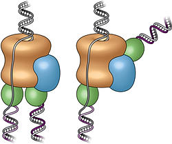
"These new images show the fully assembled and fully activated 'helicase' protein complex—which encircles and separates the two strands of the DNA double helix as it passes through a central pore in the structure—and how the helicase coordinates with the two 'polymerase' enzymes that duplicate each strand to copy the genome."

Three-dimensional structure of the active DNA helicase bound to the front-end DNA polymerase (Pol epsilon). The DNA polymerase epsilon (green) sits on top rather than the bottom of the helicase.


Studying this molecular machinery, known collectively as a "replisome," and the details of its DNA-copying process can help scientists understand what happens when DNA is miscopied—a major source of mutation that can lead to cancer—or learn more about how a single cell can eventually develop into the many cell types that make up a multicellular organism. But no one has produced a real structure of a replisome at any resolution for any organism until now.

"All the textbook drawings and descriptions of how a replisome should look and work are based on biochemical and genetic studies," Li said, likening the situation to the famous parable of the three blind men trying to describe an elephant, each looking at only one part. Those textbook drawings show the helicase moving along the DNA, separating the two strands of the double helix, with two polymerases located at the back where the DNA strand is split. In this configuration, the polymerases would add nucleotides (molecules containing the complementary A, T, G, and C bases of the genetic code) to the side-by-side split ends as they move out of the helicase to form two new complete double helix DNA strands.

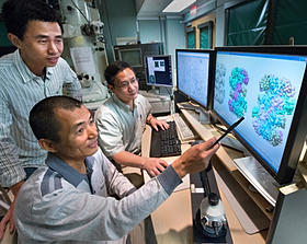
Collaborating scientists and study coauthors Zuanning Yuan, a graduate student at Stony Brook University (standing), Huilin Li of Stony Brook and Brookhaven Lab (seated, back), and Jingchuan Sun of Brookhaven Lab (seated, front) examining protein structures.

To test these assumptions, Li's group turned to the technique they had previously used to study individual components of the helicase, electron microscopy (EM). Jingchuan Sun, an EM expert in Li's lab, was essential to the success of the work. He studied samples of replisomes from baker's yeast cells—a model for all nucleus-containing cells—prepared and provided by Roxana Georgescu in Michael O'Donnell's research group at Rockefeller University. O'Donnell's group had previously published biochemical results related to this work.

"DNA replication is one of the most fundamental processes of life, so it is every biochemist's dream to see what a replisome looks like," Sun said. "Our lab has expertise and a decade of experience using electron microscopy to study DNA replication, which has prepared us well to tackle the highly mobile therefore very challenging replisome structure. Working together with the O'Donnell lab, which has done beautiful functional studies on the yeast replisome, our two groups brought perfectly complementary expertise to this project," he said.

The team's first-ever images of an intact replisome revealed that only one of the polymerases is located at the back of the helicase. The other is on the front side of the helicase, where the helicase first encounters the double-stranded helix. This means that while one of the two split DNA strands is acted on by the polymerase at the back end, the other has to thread itself back through or around the helicase to reach the front-side polymerase before having its new complementary strand assembled.

The scientists were so surprised by this finding that they asked another group at Rockefeller, led by Brian Chait, to perform additional structural studies using mass spectrometry. Yi Shi, a postdoctoral fellow in Chait's group performed this work, which confirmed the electron-microscopy-based conclusion about the unexpected architecture of the replisome.

The counterintuitive position of one polymerase at the front of the helicase suggests that it may have an unforeseen function. The authors suggest several possibilities including keeping the two "daughter" strands separate to help organize them during replication and cell division. It might also be possible that, as the single strand moves over other portions of the structure, some "surveillance" protein components check for lesions or mistakes in the nucleotide sequence before it gets copied—a sort of molecular quality control.

This architecture could also potentially play an important role in developmental biology by providing a pathway for treating the two daughter strands differently. Many modifications to DNA, including how it is packaged with other proteins, control which of the many genes in the sequence are eventually expressed in cells. An asymmetric replisome may result in asymmetric treatment of the two daughter strands during cell division—an essential step for making different tissues within a multicellular organism.

As the paper concludes, "Clearly, further studies will be required to understand the functional implications of the unexpected replisome architecture reported here."

This research was funded by the National Institutes of Health (GM103314, GM109824, GM74985, AG29979, and GM38839) and by the Howard Hughes Medical Institute.