Showing posts with label nanolithography. Show all posts

Tuesday, February 2, 2016

Nature Materials: Smallest lattice structure worldwide

The smallest lattice in the world is visible under the microscope only. Struts and braces are 0.2 µm in diameter. Total size of the lattice is about 10 µm. Photo: J. Bauer / KIT

KIT scientists now present the smallest lattice structure made by man in the Nature Materials journal. Its struts and braces are made of glassy carbon and are less than 1 µm long and 200 nm in diameter. They are smaller than comparable metamaterials by a factor of 5. The small dimension results in so far unreached ratios of strength to density. Applications as electrodes, filters or optical components might be possible. (DOI: 10.1038/nmat4561)

"Lightweight construction materials, such as bones and wood, are found everywhere in nature," Dr.-Ing. Jens Bauer of Karlsruhe Institute of Technology (KIT), the first author of the study, explains. "They have a high load-bearing capacity and small weight and, hence, serve as models for mechanical metamaterials for technical applications."

Metamaterials are materials, whose structures of some micrometers (millionths of a meter) in dimension are planned and manufactured specifically for them to possess mechanical or optical properties that cannot be reached by unstructured solids. Examples are invisibility cloaks that guide light, sound or heat around objects, materials that counterintuitively react to pressure and shear (auxetic materials) or lightweight nanomaterials of high specific stability (force per unit area and density).

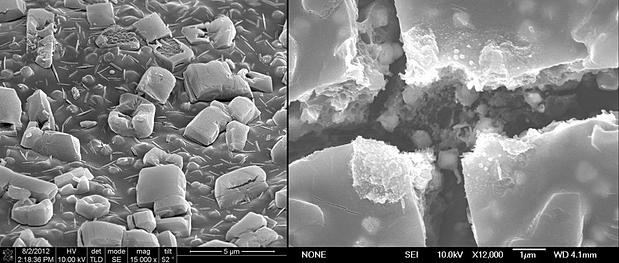
The smallest stable lattice structure worldwide presented now was produced by the established 3D laser lithography process at first. The desired structure of micrometer size is hardened in a photoresist by laser beams in a computer-controlled manner. However, resolution of this process is limited, such that struts of about 5 - 10 µm length and 1 µm in diameter can be produced only. In a subsequent step, the structure was therefore shrunk and vitrified by pyrolysis. For the first time, pyrolysis was used for manufacturing microstructured lattices. The object is exposed to temperatures of around 900°C in a vacuum furnace. As a result, chemical bonds reorient themselves. Except for carbon, all elements escape from the resist. The unordered carbon remains in the shrunk lattice structure in the form of glassy carbon. The resulting structures were tested for stability under pressure by the researchers.

"According to the results, load-bearing capacity of the lattice is very close to the theoretical limit and far above that of unstructured glassy carbon," Prof. Oliver Kraft, co-author of the study, reports. Until the end of last year, Kraft headed the Institute for Applied Materials of KIT. This year, he took over office as KIT Vice President for Research. "Diamond is the only solid having a higher specific stability."

Microstructured materials are often used for insulation or shock absorption. Open-pored materials may be used as filters in chemical industry. Metamaterials also have extraordinary optical properties that are applied in telecommunications. Glassy carbon is a high-technology material made of pure carbon. It combines glassy, ceramic properties with graphite properties and is of interest for use in electrodes of batteries or electrolysis systems.

Tuesday, June 9, 2015

Filming the film: Scientists observe photographic exposure live at the nanoscale


Advanced method opens up new opportunities for investigation of dynamic processes

Photoinduced chemical reactions are responsible for many fundamental processes and technologies, from energy conversion in nature to micro fabrication by photo-lithography. One process that is known from everyday’s life and can be observed by the naked eye, is the exposure of photographic film. At DESY's X-ray light source PETRA III, scientists have now monitored the chemical processes during a photographic exposure at the level of individual nanoscale grains in real-time. The advanced experimental method enables the investigation of a broad variety of chemical and physical processes in materials with millisecond temporal resolution, ranging from phase transitions to crystal growth. The research team lead by Prof. Jianwei (John) Miao from the University of California in Los Angeles (UCLA) and Prof. Tim Salditt from the University of Göttingen report their technique and observations in the journal Nature Materials.

The researchers investigated a photographic paper (Kodak linagraph paper Type 2167or “yellow burn paper”) that is often used to determine the position of the beam at X-ray experiments. “The photographic paper we looked at is not specially designed for X-rays. It works by changing its colour on exposure to light or X-rays,” explains DESY physicist Dr. Michael Sprung, head of the PETRA III beamline P10 where the experiments took place.

The X-rays were not only used to expose the photographic paper, but also to analyse changes of its inner composition at the same time. The paper carries a photosensitive film of a few micrometre thickness, consisting of tiny silver bromide grains dispersed in a gelatine matrix, and with an average size of about 700 nanometres. A nanometre is a millionth of a millimetre. When X-rays impinge onto such a crystalline grain, they are diffracted in a characteristic way, forming a unique pattern on the detector that reveals properties like crystal lattice spacing, chemical composition and orientation. “We could observe individual silver bromide grains within the ‘burn’ paper since the X-ray beam had a size of only 270 by 370 nanometres – smaller than the average grain,” says Salditt, who is a partner of DESY in the construction and operation of the GINIX (Göttingen Instrument for Nano-Imaging with X-Rays) at beamline P10.


The X-ray exposure starts the photolysis from silver bromide to produce silver. An absorbed X-ray photon can create many photolytic silver atoms, which grow and agglomerate at the surface and inside the silver bromide grain. The scientists observed how the silver bromide grains were strained, began to turn in the gelatine matrix and broke up into smaller crystallites as well as the growth of pure silver nano grains. The exceptionally bright beam of PETRA III together with a high-speed detector enabled the ‘filming’ of the process with up to five milliseconds temporal resolution. “We observed, for the first time, grain rotation and lattice deformation during photoinduced chemical reactions,” emphasises Miao. “We were actually surprised how fast some of these single grains rotate,” adds Sprung. “Some spin almost one time every two seconds.”

“As advanced synchrotron light sources are currently under rapid development in the US, Europe and Asia,” the authors anticipate that “in situ X-ray nanodiffraction, which enables to measure atomic resolution diffraction patterns with several millisecond temporal resolution, can be broadly applied to investigate phase transitions, chemical reactions, crystal growth, grain boundary dynamics, lattice expansion, and contraction in materials science, nanoscience, physics, and chemistry.”


Reference:
Grain rotation and lattice deformation during photoinduced chemical reactions revealed by in situ X-ray nanodiffraction; Zhifeng Huang, Matthias Bartels, Rui Xu, Markus Osterhoff, Sebastian Kalbfleisch, Michael Sprung, Akihiro Suzuki, Yukio Takahashi, Thomas N. Blanton, Tim Salditt und Jianwei Miao; Nature Materials, 2015; DOI: 10.1038/NMAT4311

Thursday, October 10, 2013

Direct ‘writing’ of artificial cell membranes on graphene

Graphene emerges as a versatile new surface to assemble model cell membranes mimicking those in the human body, with potential for applications in sensors for understanding biological processes, disease detection and drug screening.

Writing in Nature Communications, researchers at The University of Manchester led by Dr Aravind Vijayaraghavan, and Dr Michael Hirtz at the Karlsruhe Institute of Technology (KIT), have demonstrated that membranes can be directly ‘written’ on to a graphene surface using a technique known as Lipid Dip-Pen Nanolithography (L-DPN).

The human body contains 100 trillion cells, each of which is enveloped in a cell membrane which is essentially a phospholipid bi-layer membrane. These cell membranes have a plethora of proteins, ion channels and other molecules embedded in them, each performing vital functions.

It is essential, therefore, to study and understand these systems, thereby enabling their application in areas such as bio-sensing, bio-catalysis and drug-delivery. Considering that it is difficult to accomplish this by studying live cells inside the human body, scientists have developed model cell membranes on surfaces outside the body, to study the systems and processes under more convenient and accessible conditions.

Dr Vijayaraghavan’s team at Manchester and their collaborators at KIT have shown that graphene is an exciting new surface on which to assemble these model membranes, and brings many advantages compared to existing surfaces.

Dr Vijayaraghavan  said: “Firstly, the lipids spread uniformly on graphene to form high-quality membranes. Graphene has unique electronic properties; it is a semi-metal with tuneable conductivity.

“When the lipids contain binding sites such as the enzyme called biotin, we show that it actively binds with a protein called streptavidin. Also, when we use charged lipids, there is charge transfer from the lipids into graphene which changes the doping level in graphene. All of these together can be exploited to produce new types of graphene/lipids based bio-sensors.”

Dr. Michael Hirtz (KIT) explains the L-DPN technique: “The technique utilizes a very sharp tip with an apex in the range of several nanometers as a means to write lipid membranes onto surfaces in a way similar to what a quill pen does with ink on paper. The small size of the tip and the precision machine controlling it allows of course for much smaller patterns, smaller than cells, and even right down to the nanoscale.”

“By employing arrays of these tips multiple different mixtures of lipids can be written in parallel, allowing for sub-cellular sized patterns with diverse chemical composition.”
 

Source: http://www.manchester.ac.uk/aboutus/news/display/?id=10831