Showing posts with label gold nanoparticle. Show all posts

Thursday, January 7, 2016

A ‘printing press’ for nanoparticles

This new technique could facilitate use of gold nanoparticles in electronic, medical applications

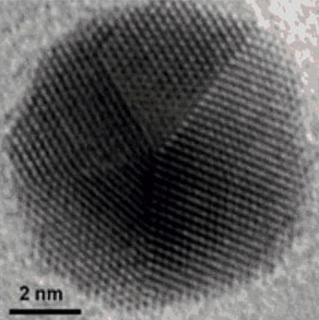
Gold nanoparticles have unusual optical, electronic and chemical properties, which scientists are seeking to put to use in a range of new technologies, from nanoelectronics to cancer treatments.

Some of the most interesting properties of nanoparticles emerge when they are brought close together – either in clusters of just a few particles or in crystals made up of millions of them.  Yet particles that are just millionths of an inch in size are too small to be manipulated by conventional lab tools, so a major challenge has been finding ways to assemble these bits of gold while controlling the three-dimensional shape of their arrangement.
One approach that researchers have developed has been to use tiny structures made from synthetic strands of DNA to help organize nanoparticles. Since DNA strands are programmed to pair with other strands in certain patterns, scientists have attached individual strands of DNA to gold particle surfaces to create a variety of assemblies. But these hybrid gold-DNA nanostructures are intricate and expensive to generate, limiting their potential for use in practical materials. The process is similar, in a sense, to producing books by hand.

Enter the nanoparticle equivalent of the printing press. It’s efficient, re-usable and carries more information than previously possible. In results reported online in Nature Chemistry, researchers from McGill’s Department of Chemistry outline a procedure for making a DNA structure with a specific pattern of strands coming out of it; at the end of each strand is a chemical “sticky patch.”  When a gold nanoparticle is brought into contact to the DNA nanostructure, it sticks to the patches. The scientists then dissolve the assembly in distilled water, separating the DNA nanostructure into its component strands and leaving behind the DNA imprint on the gold nanoparticle. (See illustration.)


“These encoded gold nanoparticles are unprecedented in their information content,” says senior author Hanadi Sleiman, who holds the Canada Research Chair in DNA Nanoscience. “The DNA nanostructures, for their part, can be re-used, much like stamps in an old printing press.”

From stained glass to optoelectronics
Some of the properties of gold nanoparticles have been recognized for centuries.  Medieval artisans added gold chloride to molten glass to create the ruby-red colour in stained-glass windows – the result, as chemists figured out much later, of the light-scattering properties of tiny gold particles. 

Now, the McGill researchers hope their new production technique will help pave the way for use of DNA-encoded nanoparticles in a range of cutting-edge technologies. First author Thomas Edwardson says the next step for the lab will be to investigate the properties of structures made from these new building blocks. “In much the same way that atoms combine to form complex molecules, patterned DNA gold particles can connect to neighbouring particles to form well-defined nanoparticle assemblies.”

These could be put to use in areas including optoelectronic nanodevices and biomedical sciences, the researchers say. The patterns of DNA strands could, for example, be engineered to target specific proteins on cancer cells, and thus serve to detect cancer or to selectively destroy cancer cells. 

Financial support for the research was provided by the Natural Sciences and Engineering Research Council of Canada, the Canada Foundation for Innovation, the Centre for Self-Assembled Chemical Structures, the Canada Research Chairs Program and the Canadian Institutes of Health Research.

“Transfer of molecular recognition information from DNA nanostructures to gold nanoparticles,” Thomas G. W. Edwardson et al, Nature Chemistry, Jan. 4, 2016. DOI: 10.1038/nchem.2420
http://www.nature.com/nchem/journal/vaop/ncurrent/full/nchem.2420.html

Tuesday, June 3, 2014

‘Quadrapeutics’ works in preclinical study of hard-to-treat tumors

Animal tests show Rice-developed technology effective against aggressive cancer

The first preclinical study of a new Rice University-developed anti-cancer technology found that a novel combination of existing clinical treatments can instantaneously detect and kill only cancer cells — often by blowing them apart — without harming surrounding normal organs. The research, which is available online this week Nature Medicine, reports that Rice’s “quadrapeutics” technology was 17 times more efficient than conventional chemoradiation therapy against aggressive, drug-resistant head and neck tumors.
The work was conducted by researchers from Rice, the University of Texas MD Anderson Cancer Center and Northeastern University.
“We address aggressive cancers that cannot be efficiently and safely treated today,” said Rice scientist Dmitri Lapotko, the study’s lead investigator. “Surgeons often cannot fully remove tumors that are intertwined with important organs. Chemotherapy and radiation are commonly used to treat the residual portions of these tumors, but some tumors become resistant to chemoradiation. Quadrapeutics steps up when standard treatments fail. At the same time, quadrapeutics complements current approaches instead of replacing them.”
quadrapeutics diagram
The first preclinical study of the anti-cancer technology "quadrapeutics" found it to be 17 times more efficient than conventional chemoradiation therapy against aggressive, drug-resistant head and neck tumors. Credit: D. Lapotko and E. Lukianova-Hleb/Rice University
Lapotko said quadrapeutics differs from other developmental cancer treatments in that it radically amplifies the intracellular effect of drugs and radiation only in cancer cells. The quadrapeutic effects are achieved by mechanical events — tiny, remotely triggered nano-explosions called “plasmonic nanobubbles.” Plasmonic nanobubbles are non-stationary vapors that expand and burst inside cancer cells in nanoseconds in response to a short, low-energy laser pulse. Plasmonic nanobubbles act as a “mechanical drug” against cancer cells that cannot be surgically removed and are otherwise resistant to radiation and chemotherapy.
In prior studies, Lapotko showed he could use plasmonic nanobubbles alone to literally blow cells apart. In quadrapeutics, his team is using them to detect and kill cancer cells in three ways. In cancer cells that survive the initial explosions, the bursting nanobubbles greatly magnify the local doses of both chemotherapy drugs and radiation. All three effects — mechanical cell destruction, intracellular drug ejection and radiation amplification — occur only in cancer cells and do not harm vital healthy cells nearby.
To administer quadrapeutics, the team uses four clinically approved components: chemotherapy drugs, radiation, near-infrared laser pulses of low energy and colloidal gold.
Dmitri Lapotko
Dmitri Lapotko
“Quadrapeutics shifts the therapeutic paradigm for cancer from materials — drugs or nanoparticles — to mechanical events that are triggered on demand only inside cancer cells,” Lapotko said. “Another strategic innovation is in complementing current macrotherapies with microtreatment. We literally bring surgery, chemotherapies and radiation therapies inside cancer cells.”
The first component of quadrapeutics is a low dose of a clinically validated chemotherapy drug. The team tested two: doxorubicin and paclitaxel. In each case, the scientists used encapsulated versions of the drug that were tagged with antibodies designed to target cancer cells. Thanks to the magnifying effect of the plasmonic nanobubbles, the intracellular dose — the amount of the drug that is active inside cancer cells — is very high even when the patient receives only a few percent of the typical clinical dose.
The second component is an injectable solution of nontoxic gold colloids, tiny spheres of gold that are thousands of times smaller than a living cell. Quadrapeutics represents a new use of colloidal gold, which has been used for decades in the clinical treatment of arthritis. In quadrapeutics, the gold colloids are tagged with cancer-specific clinically approved antibodies that cause them to accumulate and cluster together inside cancer cells. These gold “nanoclusters” do nothing until activated by a laser pulse or radiation.
Ekaterina Lukianova-Hleb
The third quadrapeutic component is a short near-infrared laser pulse that uses 1 million times less energy that a typical surgical laser. A standard endoscope delivers the laser pulse to the tumor, where the gold nanoclusters convert the laser energy into plasmonic nanobubbles.
The fourth component is a single, low dose of radiation. The gold nanoclusters amplify the deadly effects of radiation only inside cancer cells, even when the overall dose to the patient is just a few percent of the typical clinical dose.
“What kills the most-resistant cancer cells is the intracellular synergy of these components and the events we trigger in cells,” Lapotko said. “This synergy showed a 100-fold amplification of the therapeutic strength of standard chemoradiation in experiments on cancer cell cultures.”
In the Nature Medicine study, the team tested quadrapeutics against head and neck squamous cell carcinoma (HNSCC), an aggressive and lethal form of cancer that had grown resistant to both chemotherapy drugs and radiation. Quadrapeutics proved so deadly against HNSCC tumors that a single treatment using just 3 percent of the typical drug dose and 6 percent of the typical radiation dose effectively eliminated tumors in mice within one week of the administration of quadrapeutics.
Lapotko, a faculty fellow in biochemistry and cell biology and in physics and astronomy, said he is working with colleagues at MD Anderson and Northeastern to move as rapidly as possible toward prototyping and a human clinical trial. In clinical applications, quadrapeutics will be applied as either a stand-alone or intra-operative procedure using standard endoscopes and other clinical equipment and encapsulated drugs such as Doxil or Lipoplatin. Though the current study focused on head and neck tumors, Lapotko said quadrapeutics is a universal technology that can be applied for local treatment of various solid tumors, including other hard-to-treat types of brain, lung and prostate cancer. He said it might also prove especially useful for treating children due to its safety.
“The combination of aggressiveness and drug and radiation resistance is particularly problematic in tumors that cannot be fully resected, and new efficient solutions are needed,” said Dr. Ehab Hanna, a surgeon and vice chair of the Department of Head and Neck Surgery at MD Anderson, who was not involved with the testing or development of quadrapeutics. “Technologies that can merge and amplify the effects of surgery, drugs and radiation at the cellular level are ideal, and the preclinical results for quadrapeutics make it a promising candidate for clinical translation.”
Study co-authors included Rice research scientist Ekaterina Lukianova-Hleb, MD Anderson researchers Xiangwei Wu and Xiaoyang Ren and Northeastern researchers Vladimir Torchilin and Rupa Sawant.
The research was supported by the National Institutes of Health, the National Science Foundation and the Virginia and L.E. Simmons Family Foundation.
Source: http://news.rice.edu/2014/06/01/quadrapeutics-works-in-preclinical-study-of-hard-to-treat-tumors-2/#sthash.5grfIIMZ.dpuf

Saturday, April 12, 2014

Ordered arrays of nanoporous gold nanoparticles

SEM images (false color) at 25° tilt of the perfectly
ordered array of the nanoporous gold nanoparticles
formed from the 15 nm Au/30 nm Ag bilayers.
© 2012 Wang et al; licensee Beilstein-Institut.
A combination of a “top-down” approach (substrate-conformal imprint lithography) and two “bottom-up” approaches (dewetting and dealloying) enables fabrication of perfectly ordered 2-dimensional arrays of nanoporous gold nanoparticles. 

The dewetting of Au/Ag bilayers on the periodically prepatterned substrates leads to the interdiffusion of Au and Ag and the formation of an array of Au–Ag alloy nanoparticles. The array of alloy nanoparticles is transformed into an array of nanoporous gold nanoparticles by a following dealloying step. 

Large areas of this new type of material arrangement can be realized with this technique. In addition, this technique allows for the control of particle size, particle spacing, and ligament size (or pore size) by varying the period of the structure, total metal layer thickness, and the thickness ratio of the as-deposited bilayers.

Metallic nanoparticle arrays are attracting more and more attention due to their potential applications in plasmonics, magnetic memories, DNA detection, and catalytic nanowire growth. Nanoporous gold is very interesting for application in catalysi, for sensors, for actuators, and as electrodes for electrochemical supercapacitors. This is due to the unique structural, mechanical and chemical properties of this material. Nanoporous gold, already synthesized in the form of nanoparticles, possesses a much higher surface-to-volume ratio than bulk nanoporous gold films and gold nanoparticles. These nanoporous gold nanoparticles are expected to broaden the range of applications for both gold nanoparticles and nanoporous gold due to their two-level nanostructures (porosity of around 10 nm and particle size of a few hundreds of nanometers).
Solid-state dewetting of metal films is a simple “bottom-up” approach to fabricate nanoparticles. The dewetting of metal films is driven by reducing the surface energy of the film and the interface energy between the film and the substrate, and occurs by diffusion even well below the melting temperature of the film. In addition, alloy nanoparticles can be fabricated by exploiting the dewetting of metallic bilayers. By combining both, “top-down” approaches (such as lithography) and “bottom-up” approaches, an ordered array of metallic nanoparticles can be fabricated. The surface of the substrate is prepatterned into periodic structures by using laser interference lithography, focused ion beam (FIB), or substrate conformal imprint lithography (SCIL). During the dewetting of metal films onto prepatterned substrates, the periodic structure of the prepatterned substrates modulates the local excess chemical potential by the local curvature or by limiting the diffusion paths. This leads to the formation of 2-D nanoparticle arrays with well-defined particle size and particle spacing. Dealloying is a “bottom-up” approach to fabricate nanoporous gold by selectively removing or leaching the element Ag from the Au–Ag alloy in an Ag-corrosive environment. In this paper, perfectly ordered arrays of nanoporous gold nanoparticles are fabricated by using a combination of a “top-down” approach (SCIL) and two “bottom-up” approaches (dewetting and dealloying).

Full paper: http://www.beilstein-journals.org/bjnano/single/articleFullText.htm?publicId=2190-4286-3-74

Friday, October 25, 2013

Gold nanoparticles give an edge in recycling CO2

It’s a 21st-century alchemist’s dream: turning Earth’s superabundance of carbon dioxide — a greenhouse gas — into fuel or useful industrial chemicals. Researchers from Brown have shown that finely tuned gold nanoparticles can do the job. The key is maximizing the particles’ long edges, which are the active sites for the reaction.
PROVIDENCE, R.I. [Brown University] — By tuning gold nanoparticles to just the right size, researchers from Brown University have developed a catalyst that selectively converts carbon dioxide (CO2) to carbon monoxide (CO), an active carbon molecule that can be used to make alternative fuels and commodity chemicals.
“Our study shows potential of carefully designed gold nanoparticles to recycle CO2 into useful forms of carbon,” said Shouheng Sun, professor of chemistry and one of the study’s senior authors. “The work we’ve done here is preliminary, but we think there’s great potential for this technology to be scaled up for commercial applications.”
The findings are published in the Journal of the American Chemical Society.
The idea of recycling CO2 — a greenhouse gas the planet current has in excess — is enticing, but there are obstacles. CO2 is an extremely stable molecule that must be reduced to an active form like CO to make it useful. CO is used to make synthetic natural gas, methanol, and other alternative fuels.
Converting CO2 to CO isn’t easy. Prior research has shown that catalysts made of gold foil are active for this conversion, but they don’t do the job efficiently. The gold tends to react both with the CO2 and with the water in which the CO2 is dissolved, creating hydrogen byproduct rather than the desired CO.
The Brown experimental group, led by Sun and Wenlei Zhu, a graduate student in Sun’s group, wanted to see if shrinking the gold down to nanoparticles might make it more selective for CO2. They found that the nanoparticles were indeed more selective, but that the exact size of those particles was important. Eight nanometer particles had the best selectivity, achieving a 90-percent rate of conversion from CO2 to CO. Other sizes the team tested — four, six, and 10 nanometers — didn’t perform nearly as well.
“At first, that result was confusing,” said Andrew Peterson, professor of engineering and also a senior author on the paper. “As we made the particles smaller we got more activity, but when we went smaller than eight nanometers, we got less activity.”
To understand what was happening, Peterson and postdoctoral researcher Ronald Michalsky used a modeling method called density functional theory. They were able to show that the shapes of the particles at different sizes influenced their catalytic properties.
“When you take a sphere and you reduce it to smaller and smaller sizes, you tend to get many more irregular features — flat surfaces, edges and corners,” Peterson said. “What we were able to figure out is that the most active sites for converting CO2 to CO are the edge sites, while the corner sites predominantly give the by-product, which is hydrogen. So as you shrink these particles down, you’ll hit a point where you start to optimize the activity because you have a high number of these edge sites but still a low number of these corner sites. But if you go too small, the edges start to shrink and you’re left with just corners.”
Now that they understand exactly what part of the catalyst is active, the researchers are working to further optimize the particles. “There’s still a lot of room for improvement,” Peterson said. “We’re working on new particles that maximize these active sites.”
The researchers believe these findings could be an important new avenue for recycling CO2 on a commercial scale.
“Because we’re using nanoparticles, we’re using a lot less gold than in a bulk metal catalyst,” Sun said. “That lowers the cost for making such a catalyst and gives the potential to scale up.”
The work was funded by a National Science Foundation grant to the Brown-Yale Center for Chemical Innovation (CCI), which looks for ways to use CO2 as a sustainable feedstock for large-scale commodity chemicals. Other authors on the paper were Önder Metin, Haifeng Lv, Shaojun Guo, Christopher Wright, and Xiaolian Sun.

Friday, August 23, 2013

The gold standard for cell penetration

Gold nanoparticles with special coatings can deliver drugs or biosensors to a cell’s interior without damaging it.

Cells are very good at protecting their precious contents — and as a result, it’s very difficult to penetrate their membrane walls to deliver drugs, nutrients or biosensors without damaging or destroying the cell. One effective way of doing so, discovered in 2008, is to use nanoparticles of pure gold, coated with a thin layer of a special polymer. But nobody knew exactly why this combination worked so well, or how it made it through the cell wall.

Now, researchers at MIT and the Ecole Polytechnique de Lausanne in Switzerland have figured out how the process works, and the limits on the sizes of particles that can be used. Their analysis appears in the journal Nano Lettersin a paper by graduate students Reid Van Lehn, Prabhani Atukorale, Yu-Sang Yang and Randy Carney and professors Alfredo Alexander-Katz, Darrell Irvine and Francesco Stellacci.

Until now, says Van Lehn, the paper’s lead author, “the mechanism was unknown. … In this work, we wanted to simplify the process and understand the forces” that allow gold nanoparticles to penetrate cell walls without permanently damaging the membranes or rupturing the cells. The researchers did so through a combination of lab experiments and computer simulations.

The team demonstrated that the crucial first step in the process is for coated gold nanoparticles to fuse with the lipids — a category of natural fats, waxes and vitamins — that form the cell wall. The scientists also demonstrated an upper limit on the size of such particles that can penetrate the cell wall — a limit that depends on the composition of the particle’s coating.

The coating applied to the gold particles consists of a mix of hydrophobic and hydrophilic components that form a monolayer — a layer just one molecule thick — on the particle’s surface. Any of several different compounds can be used, the researchers explain.

“Cells tend to engulf things on the surface,” says Alexander-Katz, an associate professor of materials science and engineering at MIT, but it’s “very unusual” for materials to cross that membrane into the cell’s interior without causing major damage. Irvine and Stellacci demonstrated in 2008 that monolayer-coated gold nanoparticles could do so; they have since been working to better understand why and how that works.

Since the nanoparticles themselves are completely coated, the fact that they are made of gold doesn’t have any direct effect, except that gold nanoparticles are an easily prepared model system, the researchers say. However, there is some evidence that the gold particles have therapeutic properties, which could be a side benefit. 

Gold particles are also very good at capturing X-rays — so if they could be made to penetrate cancer cells, and were then heated by a beam of X-rays, they could destroy those cells from within. “So the fact that it’s gold may be useful,” says Irvine, a professor of materials science and engineering and biological engineering and member of the 
Koch Institute for Integrative Cancer Research.

Significantly, the mechanism that allows the nanoparticles to pass through the membrane seems also to seal the opening as soon as the particle has passed. “They would go through without allowing even small molecules to leak through behind them,” Van Lehn says.

Irvine says that his lab is also interested in harnessing this cell-penetrating mechanism as a way of delivering drugs to the cell’s interior, by binding them to the surface coating material. One important step in making that a useful process, he says, is finding ways to allow the nanoparticle coatings to be selective about what types of cells they attach to. “If it’s all cells, that’s not very useful,” he says, but if the coatings can be targeted to a particular cell type that is the target of a drug, that could be a significant benefit.

Another potential application of this work could be in attaching or inserting biosensing molecules on or into certain cells, Van Lehn says. In this way, scientists could detect or monitor specific biochemical markers, such as proteins that indicate the onset or decline of a disease or a metabolic process.

In general, attachment to nanoparticles’ surface coatings could provide a key to cells’ interiors for “molecules that normally wouldn’t have any ability to get through the cell membrane,” Irvine says.

Vince Rotello, a professor of chemistry at the University of Massachusetts at Amherst who was not involved in this research, says this work is “careful, well thought out and elegantly presented.” He adds, “This study provides a very interesting alternative mechanism to cell uptake of nanomaterials that could open up new therapeutic pathways.”

http://web.mit.edu/newsoffice/2013/the-gold-standard-for-cell-penetration-0823.html