Monday, April 7, 2014

Fungi Discovered In The Amazon Will Eat Your Plastic

Plastic is perhaps one of the hugest environmental problems facing Humankind and virtually all animal species around the globe. Being well known for it’s low weight, every year over 300 million tons of plastic are produced in the planet. Estimations report more plastic produced in the first decade of the 21st century, than the entire amount produced in the last century.

Only in our planet oceans, around 46.000 pieces of plastic debris, known as nurdles, are floating in each square mile, killing up to 1 million sea birds 100 thousand sea mammals adding to an unknown number of countless fish, each year.
Moreover, as far as we Humans are concerned, bisphenol A, more known as BPA, the main dioxin released by plastic containers into food and bottled water, as reached a detectable level in 93% of the people in developed countries. BPA has been long ago proven to be a carcinogen element to the Human body. Not only that, also BPA’s are proven to induce cardiovascular disease, age and puberty, obesity and developmental disorders.
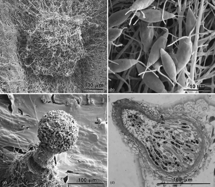
Primary fruiting structures of Pestalotiopsis microspora and Pestalosphaeria hansenii: (a) an acervulus of Pestalotiopsis microspora; (b) appendage-bearing conidiospores of Pestalotiopsis microspora; (c) a perithicium of Pestalosphaeria hansenii with an agglutinated mass of ascospores; (d) the asci of Pestalosphaeria hansenii located within a perithecium.
Due to it’s low rate of biodegradability, known to be able to take up to thousand of years to succeed, the amount of landfill goes up to 25% of all worldwide waste deposits, being from far the most comment element in them.
Being on the top of the environmental issues, a University of Yale team has recently performed a research on plastic eating fungi, and found a mushroom creating fungus able to bio-degrade polymer polyurethan, one of the most common type of worldwide produced plastic.
The Amazon is home to more species than almost anywhere else on earth. One of them, carried home recently by a group from Yale University, appears to be quite happy eating plastic in airless landfills.
The group of students, part of Yale’s annual Rainforest Expedition and Laboratory with molecular biochemistry professor Scott Strobel, ventured to the jungles of Ecuador. The mission was to allow "students to experience the scientific inquiry process in a comprehensive and creative way." The group searched for plants, and then cultured the microorganisms within the plant tissue. As it turns out, they brought back a fungus new to science with a voracious appetite for a global waste problem: polyurethane.
The fungi, Pestalotiopsis microspora, is the first anyone has found to survive on a steady diet of polyurethane alone and--even more surprising--do this in an anaerobic (oxygen-free) environment that is close to the condition at the bottom of a landfill.
Student Pria Anand recorded the microbe’s remarkable behavior and Jonathan Russell isolated the enzymes that allow the organism to degrade plastic as its food source. The Yale team published their findings in the journal Applied and Environmental Microbiologylate last year concluding the microbe is "a promising source of biodiversity from which to screen for metabolic properties useful for bioremediation." In the future, our trash compactors may simply be giant fields of voracious fungi.
Who knows what the students in the rainforest will turn up next?
http://aem.asm.org/content/77/17/6076

Organic Solar Cells More Efficient With Molecules Face-to-Face

Molecules in face-on orientation inside
organic solar cell. 
Artist: Peter Allen.
New research from North Carolina State University and UNC-Chapel Hill reveals that energy is transferred more efficiently inside of complex, three-dimensional organic solar cells when the donor molecules align face-on, rather than edge-on, relative to the acceptor. This finding may aid in the design and manufacture of more efficient and economically viable organic solar cell technology.
Organic solar cell efficiency depends upon the ease with which an exciton – the energy particle created when light is absorbed by the material – can find the interface between the donor and acceptor molecules within the cell. At the interface, the exciton is converted into charges that travel to the electrodes, creating power. While this sounds straightforward enough, the reality is that molecules within the donor and acceptor layers can mix, cluster into domains, or both, leading to variances in domain purity and size which can affect the power conversion process. Moreover, the donor and acceptor molecules have different shapes, and the way they are oriented relative to one another matters. This complexity makes it very difficult to measure the important characteristics of their structure.
NC State physicist Harald Ade, UNC-Chapel Hill chemist Wei You and collaborators from both institutions studied the molecular composition of solar cells in order to determine what aspects of the structures have the most impact on efficiency. In this project the team used advanced soft X-ray techniques to describe the orientation of molecules within the donor and acceptor materials. By manipulating this orientation in different solar cell polymers, they were able to show that a face-on alignment between donor and acceptor was much more efficient in generating power than an edge-on alignment.
“A face-on orientation is thought to allow favorable interactions for charge transfer and inhibit recombination, or charge loss, in organic solar cells,” Ade says, “though precisely what happens on the molecular level is still unclear.
“Donor and acceptor layers don’t just lie flat against each other,” Ade explains. “There’s a lot of mixing going on at the molecular level. Picture a bowl of flat pasta, like fettucine, as the donor polymer, and then add ‘ground meat,’ or a round acceptor molecule, and stir it all together. That’s your solar cell. What we want to measure, and what matters in terms of efficiency, is whether the flat part of the fettuccine hugs the round pieces of meat – a face-on orientation – or if the fettuccine is more randomly oriented, or worst case, only the narrow edges of stacked up pasta touch the meat in an edge-on orientation. It’s a complicated problem.
“This research gives us a method for measuring this molecular orientation, and will allow us to find out what the effects of orientation are and how orientation can be fine-tuned or controlled.”
The paper appears online April 6 in Nature Photonics. Fellow NC State collaborators were John Tumbleston, Brian Collins, Eliot Gann, and Wei Ma. Liqiang Yang and Andrew Stuart from UNC-Chapel Hill also contributed to the work. The work was funded by the U.S. Department of Energy, Office of Science, Basic Energy Science, the Office of Naval Research, and the National Science Foundation.
-peake-
 Note to editors: Abstract of the paper follows.
“The influence of molecular orientation on organic bulk heterojunction solar cells”
Authors: John R. Tumbleston, Brian A. Collins, Eliot Gann, Wei Ma and Harald Ade, North Carolina State University; Liqiang Yang, Andrew C. Stuart and Wei You, University of North Carolina at Chapel Hill
Published: April 6, 2014, in Nature Photonics
Abstract:
In bulk heterojunction organic photovoltaics, electron-donating and electron-accepting materials form a distributed network of heterointerfaces in the photoactive layer, where critical photo-physical processes occur. However, little is known about the structural properties of these interfaces due to their complex three-dimensional arrangement and the lack of techniques to measure local order. Here, we report that molecular orientation relative to donor/acceptor heterojunctions is an important parameter in realizing high-performance fullerene-based, bulk heterojunction solar cells. Using resonant soft X-ray scattering, we characterize the degree of molecular orientation, an order parameter that describes face-on (+1) or edge-on (-1) orientations relative to these heterointerfaces. By manipulating the degree of molecular orientation through the choice of molecular chemistry and the characteristics of the processing solvent, we are able to show the importance of this structural parameter on the performance of bulk heterojunction organic photovoltaic devices featuring the electron-donating polymers PNDT–DTBT, PBnDT–DTBT or PBnDT–TAZ.
http://news.ncsu.edu/releases/tp-adephotonics/

Self-Assembled Silver Superlattices Create Molecular Machines with Hydrogen-Bond “Hinges” and Moving “Gears”

Figure shows the arrangement of nanoparticles in
two neighboring layers of the superlattice, with
configurations on the left corresponding to the
equilibrium state of the superlattice at ambient
conditions, and the ones on the right recorded at the
end of the volume compression process. Comparison
of the configurations reveals flexure of the ligands
and gear-like rotations of the nanoparticles, with
the hydrogen-bonds between ligands anchored to
adjacent nanoparticles serving as “molecular hinges.”
(Image courtesy of Uzi Landman)
A combined computational and experimental study of self-assembled silver-based structures known as superlattices has revealed an unusual and unexpected behavior: arrays of gear-like molecular-scale machines that rotate in unison when pressure is applied to them.

Computational and experimental studies show that the superlattice structures, which are self-assembled from smaller clusters of silver nanoparticles and organic protecting molecules, form in layers with the hydrogen bonds between their components serving as “hinges” to facilitate the rotation. Movement of the “gears” is related to another unusual property of the material: increased pressure on the superlattice softens it, allowing subsequent compression to be done with significantly less force.
Materials containing the gear-like nanoparticles – each composed of nearly 500 atoms – might be useful for molecular-scale switching, sensing and even energy absorption. The complex superlattice structure is believed to be among the largest solids ever mapped in detail using a combined X-ray and computational techniques.
“As we squeeze on this material, it gets softer and softer and suddenly experiences a dramatic change,” said Uzi Landman, a Regents’ and F.E. Callaway professor in the School of Physics at the Georgia Institute of Technology. “When we look at the orientation of the microscopic structure of the crystal in the region of this transition, we see that something very unusual happens. The structures start to rotate with respect to one another, creating a molecular machine with some of the smallest moving elements ever observed.”
The gears rotate as much as 23 degrees, and return to their original position when the pressure is released. Gears in alternating layers move in opposite directions, said Landman, who is director of the Center for Computational Materials Science at Georgia Tech.
Supported by the Air Force Office of Scientific Research and the Office of Basic Energy Sciences in the Department of Energy, the research was reported April 6 in the journal Nature Materials. Researchers from Georgia Tech and the University of Toledo collaborated on the project.
The research studied superlattice structures composed of clusters with cores of 44 silver atoms each. The silver clusters are protected by 30 ligand molecules of an organic material – mercaptobenzoic acid (p-MBA) – that includes an acid group. The organic molecules are attached to the silver by sulfur atoms.
“It’s not the individual atoms that form the superlattice,” explained Landman. “You actually make the larger structure from clusters that are already crystallized. You can make an ordered array from those.”
In solution, the clusters assemble themselves into the larger superlattice, guided by the hydrogen bonds, which can only form between the p-MBA molecules at certain angles.
“The self-assembly process is guided by the desire to form hydrogen bonds,” Landman explained. “These bonds are directional and cannot vary significantly, which restricts the orientation that the molecules can have.”
The superlattice was studied first using quantum-mechanical molecular dynamics simulations conducted in Landman’s lab. The system was also studied experimentally by a research group headed by Terry Bigioni, an associate professor in the Department of Chemistry and Biochemistry at the University of Toledo.
The unusual behavior occurred as the superlattice was being compressed using hydrostatic techniques. After the structure had been compressed by about six percent of its volume, the pressure required for additional compression suddenly dropped significantly. The researchers discovered that the drop occurred when the nanocrystal components rotated, layer-by-layer, in opposite directions.
Just as the hydrogen bonds direct how the superlattice structure is formed, so also do they guide how the structure moves under pressure.
“The hydrogen bond likes to have directionality in its orientation,” Landman explained. “When you press on the superlattice, it wants to maintain the hydrogen bonds. In the process of trying to maintain the hydrogen bonds, all the organic ligands bend the silver cores in one layer one way, and those in the next layer bend and rotate the other way.”
When the nanoclusters move, the structure pivots about the hydrogen bonds, which act as “molecular hinges” to allow the rotation. The compression is possible at all, Landman noted, because the crystalline structure has about half of its space open.
The movement of the silver nanocrystallites could allow the superlattice material to serve as an energy-absorbing structure, converting force to mechanical motion. By changing the conductive properties of the silver superlattice, compressing the material could also allow it be used as molecular-scale sensors and switches.  
The combined experimental and computation study makes the silver superlattice one of the most thoroughly studied materials in the world.
“We now have complete control over a unique material that by its composition has a diversity of molecules,” Landman said. “It has metal, it has organic materials and it has a stiff metallic core surrounded by a soft material.”
For the future, the researchers plan additional experiments to learn more about the unique properties of the superlattice system. The unique system shows how unusual properties can arise when nanometer-scale systems are combined with many other small-scale units.
“We make the small particles, and they are different because small is different,” said Landman. “When you put them together, having more of them is different because that allows them to behave collectively, and that collective activity makes the difference.”
In addition to those already mentioned, Georgia Tech co-authors included research scientist Bokwon Yoon – the paper’s first author – and senior research scientists W.David Luedtke, Robert Barnett and Jianping Gao. Co-authors from the University of Toledo include Anil Desireddy and Brian E. Conn.
This research was supported by the Air Force Office of Scientific Research (AFOSR), and by the Office of Basic Energy Sciences of the U.S. Department of Energy (DOE) under Contract FG05-86ER45234. Any conclusions or opinions expressed are those of the authors and do not necessarily represent the official views of the AFOSR or the DOE.
CITATION: Bokwon Yoon, et al., “Hydrogen-bonded structure and mechanical chiral response of a silver nanoparticle superlattice.” (Nature Materials, 2014). http://dx.doi.org/ 10.1038/NMAT3923.
http://www.news.gatech.edu/2014/04/06/self-assembled-silver-superlattices-create-molecular-machines-hydrogen-bond-%E2%80%9Chinges%E2%80%9D-and

Thursday, April 3, 2014

Quantum Photon Properties Revealed in Another Particle—the Plasmon

For years, researchers have been interested in developing quantum computers—the theoretical next generation of technology that will outperform conventional computers. Instead of holding data in bits, the digital units used by computers today, quantum computers store information in units called "qubits." One approach for computing with qubits relies on the creation of two single photons that interfere with one another in a device called a waveguide. Results from a recent applied science study at Caltech support the idea that waveguides coupled with another quantum particle—the surface plasmon—could also become an important piece of the quantum computing puzzle.

The work was published in the print version of the journal Nature Photonics the week of March 31.

As their name suggests, surface plasmons exist on a surface—in this case the surface of a metal, at the point where the metal meets the air. Metals are conductive materials, which means that electrons within the metal are free to move around. On the surface of the metal, these free electrons move together, in a collective motion, creating waves of electrons. Plasmons—the quantum particles of these coordinated waves—are akin to photons, the quantum particles of light (and all other forms of electromagnetic radiation).

"If you imagine the surface of a metal is like a sea of electrons, then surface plasmons are the ripples or waves on this sea," says graduate student Jim Fakonas, first author on the study.
These waves are especially interesting because they oscillate at optical frequencies. Therefore, if you shine a light at the metal surface, you can launch one of these plasmon waves, pushing the ripples of electrons across the surface of the metal. Because these plasmons directly couple with light, researchers have used them in photovoltaic cells and other applications for solar energy. In the future, they may also hold promise for applications in quantum computing.

However, the plasmon's odd behavior, which falls somewhere between that of an electron and that of a photon, makes it difficult to characterize. "According to quantum theory, it should be possible to analyze these plasmonic waves using quantum mechanics"—the physics that governs the behavior of matter and light at the atomic and subatomic scale—"in the same way that we can use it to study electromagnetic waves, like light," Fakonas says. However, in the past, researchers were lacking the experimental evidence to support this theory.

To find that evidence, Fakonas and his colleagues in the laboratory of Harry Atwater, Howard Hughes Professor of Applied Physics and Materials Science, looked at one particular phenomenon observed of photons—quantum interference—to see if plasmons also exhibit this effect.

The applied scientists borrowed their experimental technique from a classic test of quantum interference in which two single, identical photons are launched at one another through opposite sides of a 50/50 beam splitter, a device that acts as an imperfect mirror, reflecting half of the light that reaches its surface while allowing the the other half of the light to pass through. If quantum interference is observed, both identical photons must emerge together on the same side of the beam splitter, with their presence confirmed by photon detectors on both sides of the mirror.

Since plasmons are not exactly like photons, they cannot be used in mirrored optical beam splitters. Therefore, to test for quantum interference in plasmons, Fakonas and his colleagues made two waveguide paths for the plasmons on the surface of a tiny silicon chip. Because plasmons are very lossy—that is, easily absorbed into materials that surround them—the path is kept short, contained within a 10-micron-square chip, which reduces absorption along the way.

The waveguides, which together form a device called a directional coupler, act as a functional equivalent to a 50/50 beam splitter, directing the paths of the two plasmons to interfere with one another. The plasmons can exit the waveguides at one of two output paths that are each observed by a detector; if both plasmons exit the directional coupler together—meaning that quantum interference is observed—the pair of plasmons will only set off one of the two detectors.

Indeed, the experiment confirmed that two indistinguishable photons can be converted into two indistinguishable surface plasmons that, like photons, display quantum interference.
This finding could be important for the development of quantum computing, says Atwater. "Remarkably, plasmons are coherent enough to exhibit quantum interference in waveguides," he says. "These plasmon waveguides can be integrated in compact chip-based devices and circuits, which may one day enable computation and measurement schemes based on quantum interference."

Before this experiment, some researchers wondered if the photon–metal interaction necessary to create a surface plasmon would prevent the plasmons from exhibiting quantum interference. "Our experiment shows this is not a concern," Fakonas says.
"We learned something new about the quantum mechanics of surface plasmons. The main thing is that we were able to validate the theoretical prediction; we showed that this type of interference is possible with plasmons, and we did a pretty clean measurement," he says. "The quantum interference displayed by plasmons appeared to be almost identical to that of photons, so I think it would be very difficult for someone to design a different structure that would improve upon this result."

The work was published in a paper titled "Two-plasmon quantum interference." In addition to Fakonas and Atwater, the other coauthors are Caltech undergraduate Hyunseok Lee and former undergraduate Yousif A. Kelaita (BS '12). The work was supported by funding from the Air Force Office of Scientific Research, and the waveguide was fabricated at the Kavli Nanoscience Institute at Caltech.

Source: http://www.caltech.edu/content/quantum-photon-properties-revealed-another-particle-plasmon#sthash.70Ss0F6E.dpuf

Nanoparticles cause cancer cells to self-destruct

Using magnetically controlled nanoparticles to force tumour cells to ‘self-destruct’ sounds like science fiction, but could be a future part of cancer treatment, according to research from Lund University in Sweden.
“The clever thing about the technique is that we can target selected cells without harming surrounding tissue. There are many ways to kill cells, but this method is contained and remote-controlled”, said Professor Erik Renström.
The point of the new technique is that it is much more targeted than trying to kill cancer cells with techniques such as chemotherapy.
“Chemotherapy can also affect healthy cells in the body, and it therefore has serious side-effects. Radiotherapy can also affect healthy tissue around the tumour.
“Our technique, on the other hand, is able to attack only the tumour cells”, said Enming Zhang, one of the first authors of the study.
In brief, the technique involves getting the nanoparticles into a tumour cell, where they bind to lysosomes, the units in the cell that perform ‘cleaning patrols’. The lysosomes have the ability to break down foreign substances that have entered a cell. They can also break down the entire cell through a process known as ‘controlled cell death’, a type of destruction where damaged cells dissolve themselves.
The researchers have used nanoparticles of iron oxide that have been treated with a special form of magnetism. Once the particles are inside the cancer cells, the cells are exposed to a magnetic field, and the nanoparticles begin to rotate in a way that causes the lysosomes to start destroying the cells.
The research group at Lund University is not the first to try and treat cancer using supermagnetic nanoparticles. However, previous attempts have focused on using the magnetic field to create heat that kills the cancer cells. The problem with this is that the heat can cause inflammation that risks harming surrounding, healthy tissue. The new method, on the other hand, in which the rotation of the magnetic nanoparticles can be controlled, only affects the tumour cells that the nanoparticles have entered.
The new technique is primarily intended for cancer treatment, but according to Erik Renström and his colleague Enming Zhang there may be other areas of application. One example is autoimmune diseases such as type 1 diabetes, in which the immune system attacks the body’s own insulin production.
The ‘superparamagnetic nanoparticles’ have attracted a lot of interest from academia and industry in recent years. They are being tested in research on new diagnostic laboratory tests, new methods of viewing phenomena in living tissue, and new drugs.
The researchers at Lund University have a patent pending for their technique with the rotating nanoparticles. However, a lot of work remains before it can be transferred from the laboratory to clinical trials on patients.
The study is a collaboration between physicists, chemists, engineers and doctors from Sweden, Germany and the USA. It has been published in the American journal ACS Nano.
Source: http://www.lunduniversity.lu.se/o.o.i.s?id=24890&news_item=6140