Showing posts with label fundamental physics. Show all posts

Friday, June 17, 2016

Physicists measured something new in the radioactive decay of neutrons.


The experiment inspired theorists; future ones could reveal new physics.

A physics experiment performed at the National Institute of Standards and Technology (NIST) has enhanced scientists’ understanding of how free neutrons decay into other particles. The work provides the first measurement of the energy spectrum of photons, or particles of light, that are released in the otherwise extensively measured process known as neutron beta decay. The details of this decay process are important because, for example, they help to explain the observed amounts of hydrogen and other light atoms created just after the Big Bang.

Published in Physical Review Letters, the findings confirm physicists’ big-picture understanding of the way particles and forces work together in the universe—an understanding known as the Standard Model. The work has stimulated new theoretical activity in quantum electrodynamics (QED), the modern theory of how matter interacts with light. The team’s approach could also help search for new physics that lies beyond the Standard Model.

Neutrons are well known as one of the three kinds of particles that form atoms. Present in all atoms except the most common form of hydrogen, neutrons together with protons form the atomic nucleus. However, “free” neutrons not bound within a nucleus decay in about 15 minutes on average. Most frequently, a neutron transforms through the beta decay process into a proton, an electron, a photon, and the antimatter version of the neutrino, an abundant but elusive particle that rarely interacts with matter.

The photons from beta decay are what the research team wanted to explore. These photons have a range of possible energies predicted by QED, which has worked very well as a theory for decades. But no one had actually checked this aspect of QED with high precision.

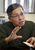
“We weren’t expecting to see anything unusual,” said NIST physicist Jeff Nico, “but we wanted to test QED’s predictions very precisely in a way no one has done before.”

Nico and his colleagues, who represent nine research institutions, performed their measurements at the NIST Center for Neutron Research (NCNR). It produces an intense beam of slow-moving neutrons whose photon emissions can be detected with the same setup used for earlier precision measurements of the neutron’s lifetime.

The team measured two aspects of neutron decay: the energy spectrum of the photons, and also its branching ratio, which can provide information on how frequently the decays were accompanied by photons above a specific energy. The results of this effort gave them a branching ratio measurement more than twice as accurate as the previous value, and the first measurement of the energy spectrum.

“Everything we found was consistent with the predominant QED calculations,” Nico said. “We got quite a good match with theory on the energy spectrum, and we reduced the uncertainty in the branching ratio.”

According to Nico, the results provided specific information that theoretical physicists are already using to further develop QED to provide more detailed descriptions of neutron beta decay.

The results serve as a needed check on the Standard Model, said Nico, and validates the team’s experimental approach as a way to go beyond it. With better detectors, the approach could be used to search for so-called “right-handed” neutrinos, which have not yet been detected in nature, and potential time-reversal symmetry violations, which could explain why there is much more matter than antimatter in the universe.

Paper: M.J. Bales, R. Alarcon, C.D. Bass, E.J. Beise, H. Breuer, J. Byrne, T.E. Chupp, K.J. Coakley, R.L. Cooper, M.S. Dewey, S. Gardner, T.R. Gentile, D. He, H.P. Mumm, J.S. Nico, B. O'Neill, A.K. Thompson and F.E. Wietfeldt (RDK II Collaboration). Precision measurement of the radiative beta decay of the free neutron. Physical Review Letters. June 14, 2016, DOI: 10.1103/PhysRevLett.116.242501

Tuesday, September 22, 2015

Physicists determine the three-dimensional positions of individual atoms for the first time


Atoms are the building blocks of all matter on Earth, and the patterns in which they are arranged dictate how strong, conductive or flexible a material will be. Now, scientists at UCLA have used a powerful microscope to image the three-dimensional positions of individual atoms to a precision of 19 trillionths of a meter, which is several times smaller than a hydrogen atom.

Their observations make it possible, for the first time, to infer the macroscopic properties of materials based on their structural arrangements of atoms, which will guide how scientists and engineers build aircraft components, for example. The research, led by Jianwei (John) Miao, a UCLA professor of physics and astronomy and a member of UCLA’s California NanoSystems Institute, is published Sept. 21 in the online edition of the journal Nature Materials.

For more than 100 years, researchers have inferred how atoms are arranged in three-dimensional space using a technique called X-ray crystallography, which involves measuring how light waves scatter off of a crystal. However, X-ray crystallography only yields information about the average positions of many billions of atoms in the crystal, and not about individual atoms’ precise coordinates.

“It’s like taking an average of people on Earth,” Miao said. “Most people have a head, two eyes, a nose and two ears. But an image of the average person will still look different from you and me.”
Because X-ray crystallography doesn’t reveal the structure of a material on a per-atom basis, the technique can’t identify tiny imperfections in materials such as the absence of a single atom. These imperfections, known as point defects, can weaken materials, which can be dangerous when the materials are components of machines like jet engines.

“Point defects are very important to modern science and technology,” Miao said.

Miao and his team used a technique known as scanning transmission electron microscopy, in which a beam of electrons smaller than the size of a hydrogen atom is scanned over a sample and measures how many electrons interact with the atoms at each scan position. The method reveals the atomic structure of materials because different arrangements of atoms cause electrons to interact in different ways.
However, scanning transmission electron microscopes only produce two-dimensional images. So creating a 3-D picture requires scientists to scan the sample once, tilt it by a few degrees and re-scan it — repeating the process until the desired spatial resolution is achieved — before combining the data from each scan using a computer algorithm. The downside of this technique is that the repeated electron beam radiation can progressively damage the sample.

Using a scanning transmission electron microscope at the Lawrence Berkeley National Laboratory’s Molecular Foundry, Miao and his colleagues analyzed a small piece of tungsten, an element used in incandescent light bulbs. As the sample was tilted 62 times, the researchers were able to slowly assemble a 3-D model of 3,769 atoms in the tip of the tungsten sample.
The experiment was time consuming because the researchers had to wait several minutes after each tilt for the setup to stabilize.

“Our measurements are so precise, and any vibrations — like a person walking by — can affect what we measure,” said Peter Ercius, a staff scientist at Lawrence Berkeley National Laboratory and an author of the paper.

The researchers compared the images from the first and last scans to verify that the tungsten had not been damaged by the radiation, thanks to the electron beam energy being kept below the radiation damage threshold of tungsten.

Miao and his team showed that the atoms in the tip of the tungsten sample were arranged in nine layers, the sixth of which contained a point defect. The researchers believe the defect was either a hole in an otherwise filled layer of atoms or one or more interloping atoms of a lighter element such as carbon.

Regardless of the nature of the point defect, the researchers’ ability to detect its presence is significant, demonstrating for the first time that the coordinates of individual atoms and point defects can be recorded in three dimensions.

“We made a big breakthrough,” Miao said.
Miao and his team plan to build on their results by studying how atoms are arranged in materials that possess magnetism or energy storage functions, which will help inform our understanding of the properties of these important materials at the most fundamental scale.
“I think this work will create a paradigm shift in how materials are characterized in the 21st century,” he said. “Point defects strongly influence a material’s properties and are discussed in many physics and materials science textbooks. Our results are the first experimental determination of a point defect inside a material in three dimensions.”
The study’s co-authors include Rui Xu, Chien-Chun Chen, Li Wu, Mary Scott, Matthias Bartels, Yongsoo Yang and Michael Sawaya, all of UCLA; as well as Colin Ophus of Lawrence Berkeley National Laboratory; Wolfgang Theis of the University of Birmingham; Hadi Ramezani-Dakhel and Hendrik Heinz of the University of Akron; and Laurence Marks of Northwestern University.

Tuesday, October 1, 2013

Fundamental physicists discover surprise new use for super-chilled neutrons to measure the movement of viruses.

•    First evidence that ultra-cold neutrons interact with moving nano-sized particles provides a new tool for chemists, biologists and engineers

•    Billiard-ball collisions may also explain inaccuracies in 60-year-old experiments to measure the lifetime of the neutron and explain the origin of matter in the universe

Physicists working on a 60-year-old experiment to understand the origin of matter in the universe have uncovered a new tool for studying the movement of tiny particles along a surface, such as a virus travelling along a cell membrane. The new tool utilises ultra-cold neutrons (UCNs) that move slower than most people can run and will allow scientists to map the movement of tiny objects with previously unattainable precision. This discovery, made at the Institut Laue-Langevin, the world’s flagship centre for neutron science and the home of ultra-cold neutron research for over 25 years, is published today in Crystallography Reports.

Since their discovery in 1969, UCNs have been used by experimental physicists to answer fundamental questions about the universe, such as the origin of matter and how gravity fits into the standard model of particle physics. They do this by collecting them in traps and monitoring properties such as their energies or lifetime at high precision.

However, the average storage time of the UCNs in their traps was always much lower than expected, affecting the quality of observations. In 1999 Dr Valery Nesvizhevsky and colleagues at the ILL discovered a new phenomenon that might explain these losses. They found that occasionally a UCN in the trap was given a small thermal ‘kick’. This occurred in just 1 out of every 10,000,000 collisions, but the origin of this ‘kick’ was unknown.
As other hypotheses were ruled out, Dr Nesvizhevsky started to consider the influence of the nanoparticles or nanodroplets which were known to populate a layer immediately above the surface of most materials, including those of the trap’s interior.

To tests this hypothesis Dr Nesvizhevsky and his colleagues returned to the UCN apparatus at the ILL. They placed samples with nanoparticle surface layers of known size distribution into the UCN trap and observed the interactions.
The team discovered that the change in UCN energy was induced by ‘billiard-ball-like’ collisions with moving surface nanoparticles, thus providing the first proof that these nanoparticles are not stationary.

The low energy levels of UCNs means they usually bounce off the inner walls and remain in the trap. However these shots of extra energy caused by the interaction with the nanoparticles gives them just enough energy either to overcome gravity and escape out of the top of the chamber, which is left open, or to pass right through the chamber walls.

This phenomenon has two very dramatic consequences:
  1. It could explain discrepancies in the results of 60-year-old experiments measuring the lifetime of the neutron, the results of which differ by about 10 seconds, much more than the reported uncertainties would allow. A precise figure could affect conclusions on the origin of matter in the early universe, as well as on the number of families of elementary particles existing in nature, and could modify models of star formation.
  2. It also gives science a brand new and uniquely accurate tool for studying for the very first time how nanoparticles move around and interact with material surfaces, particular through van der Waals/Casimir interactions, in all kinds of natural and man-made systems. Currently no other techniques exist to make these measurements.

Potential applications of this technique are vast and include the production of chemicals and semiconductors, catalytic converters, integrated circuits used in electronic devices and silver halide salts used in photographic films. The tool could also be used to study for the first time how biological molecules move along a surface such as viruses along a biological membrane.
For their next experiments, Valery and his colleagues have secured further beam time at the ILL and will be inviting scientists from various disciplines to provide samples from their own research for analysis with UCNs to prove the validity of this new technique.

Quotes

Dr Valery Nesvizhevsky said: “We found this brand new scientific tool by chance. We never thought UCNs might have such practical uses. The implications of these findings for fundamental physics are sure to be a hot topic and I expect there will be some debate as to how much these thermal kicks contribute to the uncertainties around the lifetime of the neutron measurements. However the potential in this new technique for studying nanoparticle dynamics is a certainty and we look forward to working with researchers from across the scientific disciplines to realise its potential.”

Dr Valentin Gordeliy is heads of the Membrane Transportation Group at the Institut de Biologie Structurale and has met with Dr Nesvizhevsky to discuss the possible application of the new ultra-cold neutron technique in his own line of research: “This is a great piece of science and if its feasibility can be verified, a potentially very exciting discovery for my own work in structural biology. In the human body there are a great many nano-scale structures, the motions of those correspond to time-scale of this technique. These include viruses, and various proteins and protein complexes including histones, the scaffolds of DNA that regulate the expression of genes, helping turning them on and off. One of the most attractive objects for study would be membrane proteins that help maintain the health of the whole body and are therefore targeted by 60% of existing drugs. Whilst other tools such as electron microscopes can be used they are not suitable to study dynamics. Also the possibility of watching the evolution of complex interactions such as that of a virus and a membrane protein would provide new insights for drug discovery as well as a better understanding of how our body functions.”