 | |
|
For the sake of brevity, only the very basic and universal concepts about microtubules and their organization into flagella will be presented here, leaving many questions unanswered. You will find that textbooks provide more complete descriptions of microtubules and their structures and functions, but they also leave many questions unanswered. Textbooks seldom tell us is how much science knows and does not know about them, and of course they cannot be up to date with the latest discoveries. To fully understand a subject it is important to go to multiple sources. If the subject is especially important to you, you should seek the primary literature, namely original research reports.
"Building blocks" of microtubules - tubulins
All eukaryotic cells produce the protein tubulin, in the usual way. The usual way, of course, is by transcription of genes coding for tubulin to produce messenger RNA, followed by the translation of mRNA by the ribosomes in order to produce protein. Cells maintain at least two types of tubulin, which we call alpha tubulin and beta tubulin. However, it is doubtful that the two types can found in cells as individual proteins.
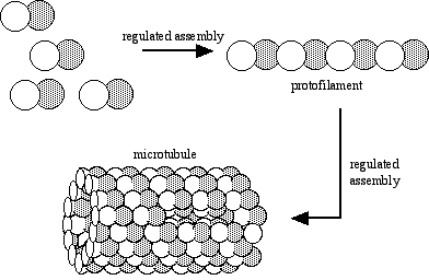
Alpha and beta tubulin spontaneously bind one another to form a functional subunit that we call a heterodimer. A heterodimer is a protein that consists of two different gene products. The term is entirely descriptive - the prefix hetero- means "different," the prefix di- means "two," and the suffix -mer refers to a unit, in this case a single polypeptide. Obviously, cells do not continue to make tubulin (or any other protein) until they run out of resources. Some process must regulate the synthesis of tubulin. A common regulatory mechanism is feedback inhibition.

The figure illustrates the inhibition of tubulin synthesis by the presence of heterodimers in the system. Exactly how that inhibition takes place is irrelevant to this discussion. More about the important concept of feedback inhibition can be found elsewhere.
Assembly of microtubules
When intracellular conditions favor assembly, tubulin heterodimers assemble into linear protofilaments. Protofilaments in turn assemble into microtubules. All such assembly is subject to regulation by the cell.
Dynamic instability of microtubules
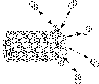
Under steady state conditions a microtubule may appear to be completely stable, however there is action taking place constantly. Populations of microtubules usually consist of some that are shrinking and some that are growing. A single microtubule can oscillate between growth and shortening phases. During growth, heterodimers are added on to the end of a microtubule, and during shrinkage they come off as intact subunits. The same heterodimer can come off and go back on.
Since even apparently stable microtubular structures have an intrinsic instability, they are considered to be in a dynamic equilibrium, or steady state. Look here to learn about the difference between a steady state and a true equilibrium.
Cilia and Flagella
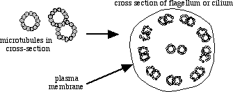
To understand the regulation of microtubule assembly and function in any organism is a difficult task. To study microtubules in cells as complex vertebrate (e.g., human) cells is a nearly impossible task, without a few "hints" as to how to proceed. The basic mechanisms can be worked out using a much less complex biological model such as a flagellate. For example, the flagella of the photosynthetic protist Chlamydomonas are composed of microtubules, as are all flagella and cilia.Cilia and flagella have the same basic structure. They are attached to structures known as basal bodies, which in turn are anchored to the cytoplasmic side of the plasma membrane. From the basal bodies the microtubule "backbone" extends, pushing the plasma membrane out with it.

The treatment given here to the structure of microtubules ignores their true complexity. Functional microtubules include associated proteins, anchoring sites and organizing centers, sites for enzyme activity, etc. In cilia and flagella, tubulin forms a core structure to which other proteins contribute structures called dynein arms, radial spokes, and nexin links. The arms, spokes, and links hold microtubules together and allow interaction between microtubules that is superficially similar to the sliding of actin and myosin filaments in muscle contraction.
Ciliary and Flagellar Motion
One might appreciate the complexity of microtubular organelles by looking at the motion of cilia and flagella. Despite the similarities in structure, the difference in nature of motility by flagella versus cilia is profound, as one can see by comparing representatives of the groups Ciliophora (the ciliates) and Mastigophora (the flagellates). Ciliates and flagellates behave differently, live in different habitats and occupy different niches, and likely represent two different evolutionary lineages. The main difference in function is in how they are organized.
Flagella are much longer than cilia and are usually present singly or in pairs. A single flagellum may propel the cell with a whip-like motion. A pair of flagella may move in a synchronized manner to pull the organism through the water, in a way similar to the breast stroke of a human swimmer.
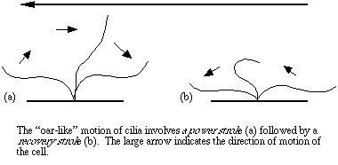
Cilia tend to cover the surface area of a cell. Both cilia and flagella bend as the microtubules slide past one another. The arrangement of cilia permits their coordinated movement in response to signals from the cytoplasm. A small ciliate may have hundreds of individual cilia, all beating in a coordinated manner. How is all of the sliding and bending coordinated? How does the organism "decide" in what direction to move, or how to turn, rotate, or feed? How does it convey the information to hundreds of cilia to bend in a certain way? Questions of that nature are fascinating to cell biologists. They are very difficult to address, because each system is so complex. Nevertheless, with a genome about a hundred times smaller than that of a human, a typical protist is much easier to study than a human cell.Flagella are much longer than cilia and are usually present singly or in pairs. A single flagellum may propel the cell with a whip-like motion. A pair of flagella may move in a synchronized manner to pull the organism through the water, in a way similar to the breast stroke of a human swimmer.

Source: http://www.ruf.rice.edu/~bioslabs/studies/invertebrates/microtubules.html

No comments:
Post a Comment